de Winter's T Waves
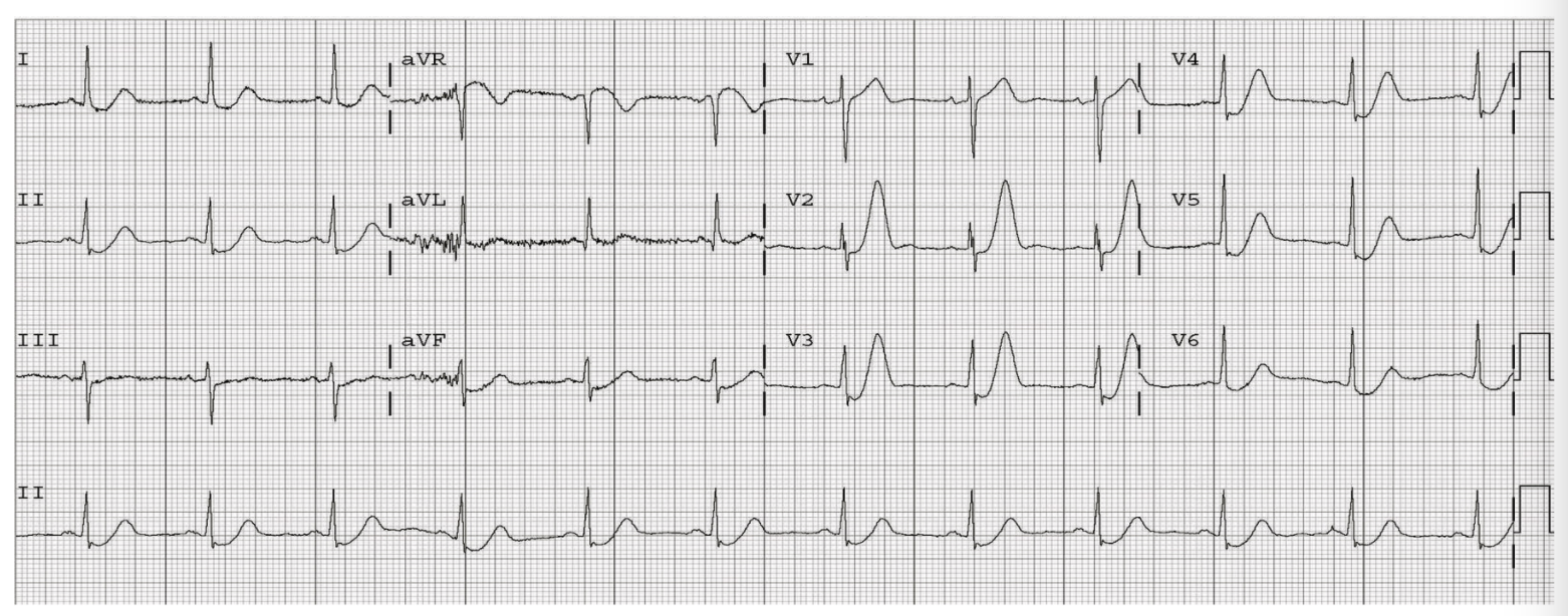
A 47 year old male presents with chest pain. His ECG is shown below. What does it show?
What is this called?
Is this a STEMI?
If you had no Cath lab, would you thrombolyse?
What is this called?
Is this a STEMI?
If you had no Cath lab, would you thrombolyse?
Answer
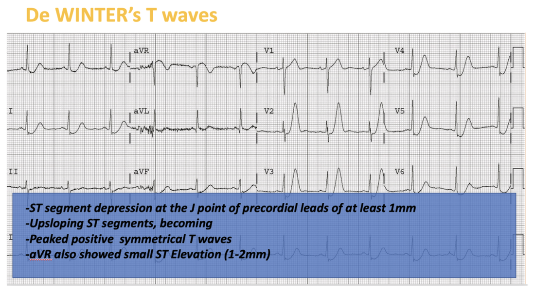
These are de Winter's T waves. They are a STEMI EQUIVALENT
De Winter’s T Waves were first described by De Winter in an article to the editor of the New England Journal of Medicine in 2008(1). They are considered an ST-elevation myocardial infarction(STEMI) equivalent and knowledge of this sign is important as ST depression is not the classic appearance of an STEMI.
Using ECG’s from a large database the authors described a new ECG pattern without ST elevation, that indicated proximal left anterior descending(LAD) coronary artery occlusion.
The pattern was seen in the precordial leads is:
It was described as a static pattern, rather than evolving with changes to the ST segments. Since then other studies have in fact shown evolution to the classic STEMI pattern does occur.
This is considered to be an anterior STEMI Equivalent.
In 2009 Verovden el al (2) duplicated these findings in patients with anterior wall myocardial infarction. The distinct ECG pattern was identified in approximately 2% of patients with LAD stenosis, in that study also. These patients were more likely to be younger and male and tended to have hypercholesterolaemia.
References
Using ECG’s from a large database the authors described a new ECG pattern without ST elevation, that indicated proximal left anterior descending(LAD) coronary artery occlusion.
The pattern was seen in the precordial leads is:
- ST segment depression at the J point of precordial leads of 1-3 mm
- Up-sloping ST segments, becoming peaked positive symmetrical T waves
- Occurred in leads V1 – V6
- QRS complexes were in some cases slightly widened
- Loss of precordial R-wave progression
- aVR also showed small ST Elevation of 1-2mm
It was described as a static pattern, rather than evolving with changes to the ST segments. Since then other studies have in fact shown evolution to the classic STEMI pattern does occur.
This is considered to be an anterior STEMI Equivalent.
In 2009 Verovden el al (2) duplicated these findings in patients with anterior wall myocardial infarction. The distinct ECG pattern was identified in approximately 2% of patients with LAD stenosis, in that study also. These patients were more likely to be younger and male and tended to have hypercholesterolaemia.
References
- De Winter Rd et al A new sign of proximal lad occlusion, NEJM 2008:359: 2071-2073 Nov 6
- Verovden NJ Et al Persistent precordial “hyperacute” T waves signify proximal left anterior descending artery occlusion. Heart, 2009 Oct; 95(2): 1701-6


