CARDIAC BOOTCAMP
  • HOME
  • Course Login
    • Log In
    • Log Out
  • SYLLABUS
  • STORE
    • Online Self Study Course
  • ECG OF THE WEEK

ECG OF THE WEEK

BECOME A COURSE MEMBER
Each Week we put up one ECG case for you....because it's easier to learn from cases.
The ECG of the week is FREE to everyone. To become an expert at ECG's join the Cardiac Bootcamp Course. It's a great way to Learn!
​Or come to one of our LIVE Cardiac Bootcamps

A 40 yo with depressed conscious state

25/5/2021

2 Comments

 
A 40 yo is brought to the emergency department with the following vitals:
  • GCS 4
  • SBP 70mmHg
  • HR 140
  • RR 50
  • T 39C
There are no signs of injury. Heart sounds are dual and no extra sounds.  There are reduced breath sounds and crepitations in the left base. The abdomen is soft and the patient appears to have peripheral cyanosis. The patient has a known history of IVDU and has been found in a chair, not rousable.
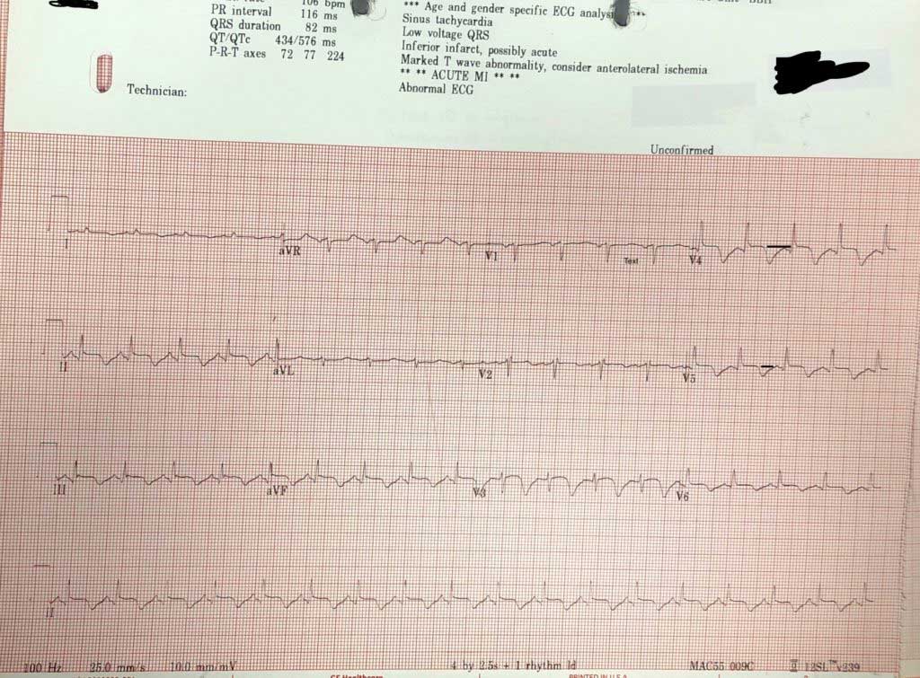
An  ECG is done.

  1. What does the ECG show?
  2. What are your differential diagnoses for this patient?
  3. What is the most likely diagnosis? 
  4. The computer says acute AMI. Is it?​​
Picture
If we use the 'ECG in 20 Seconds Approach' to describe the ECG, we get the following:
Rate/Rhythm 
106

  • Is it sinus?– P waves are upright in II and inverted in aVR; Yes
  • Is there a P for every QRS? Yes
QRS:
  • Is it tall/small?: It’s about right, there is no hypertrophy.
  • Is it wide/narrow? It’s narrow
  • Is it of abnormal morphology ie., delta wave? No
  • Is it clumped?(just incase we miss a Mobitz- but it’s not slow enough) No
ST-T
  • Remember the baseline is the T-P line
    • If we look we see that there is some ST elevation in some areas of the ECG of about 1mm, although this is difficult to gauge as the T wave goes straight into the p wave.
  • The most visible abnormality is the inverted T waves.
What causes inverted T waves?
MEMBERS Click here to read the more
2 Comments
Toyin Yusuf
26/5/2021 09:36:20 am

Normal variant in Children
Acute Myocardial ischaemia
Pulmonary embolism
Wellens angina
Raised intracranial pressure
Positive stress test .

Reply
luc de sonville
27/5/2021 02:03:17 am

infero-lateraal MI

Reply


Leave a Reply.

    Author

    Dr Peter Kas
    ​Emergency Physician

    Picture
    Sign up for
    ECG of the week
     Its free

    Archives

    July 2022
    June 2022
    March 2022
    December 2021
    November 2021
    October 2021
    September 2021
    August 2021
    July 2021
    June 2021
    May 2021
    April 2021
    March 2021
    February 2021
    January 2021
    December 2020
    November 2020
    September 2020
    August 2020
    July 2020

    Categories

    All
    Atrial Fibrillation
    Atrial Flutter
    Atrial Tachycardia
    Axis
    Bradycardia
    Cardiac Arrest
    ECG Mimics
    Lead Reversal
    Pacing
    Paediatric ECG
    Pericarditis
    QT Interval
    STEMI Mimics
    SVT
    Wellens
    Wide Complex Tachycardia

Our Other Websites to Visit

www.emcore.com.au
www.resus.com.au

Contact Us
© COPYRIGHT 2015. ALL RIGHTS RESERVED.
  • HOME
  • Course Login
    • Log In
    • Log Out
  • SYLLABUS
  • STORE
    • Online Self Study Course
  • ECG OF THE WEEK