CARDIAC BOOTCAMP
  • HOME
  • Course Login
    • Log In
    • Log Out
  • SYLLABUS
  • STORE
    • Online Self Study Course
    • Cardiac Video Course
  • ECG OF THE WEEK

ECG OF THE WEEK

BECOME A COURSE MEMBER
Each Week we put up one ECG case for you....because it's easier to learn from cases.
The ECG of the week is FREE to everyone. To become an expert at ECG's join the Cardiac Bootcamp Course. It's a great way to Learn!
​Or come to one of our LIVE Cardiac Bootcamps

A 28 yo woman with sinus tachycardia

29/6/2021

0 Comments

 
Sinus Tachycardia in the asymptomatic patient may seem benign, but..............
A 28 yo woman presents to the Emergency Department following an ankle injury. She has a  past medical history of diabetes. Her blood sugar on the day is 24 mmol/L and it's obvious, by the fact that she has just eaten a chocolate bar in the waiting room, that she may have issues with her glycaemic control. 
​She is found to be tachycardic, however has no symptoms of palpitations or chest pain or shortness of breath. The ankle injury is not causing significant pain at rest, but the tachycardia continues. She is managed for her ankle injury and discharged.
This same patient had been in the emergency department the day before for initial treatment and was also tachycardic at that time.
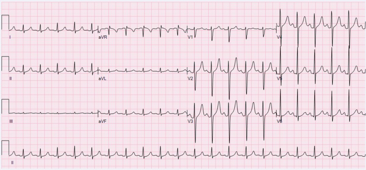
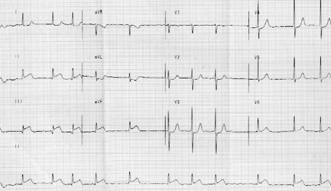
Her ECG is shown below (this is not the actual patient's ECG but one similar).
What do you think?
Picture
Please note that this is not the actual patient's ECG, but a similar ECG with Source: Wikipedia.
This is a straightforward sinus tachycardia.
Why would I have called the patient back when I saw the history?

​Persistent sinus tachycardia is worrying.
​Simple sinus tachycardia may be caused by responses to pain, drinking coffee or even eating some chocolate, We need to separate the benign from the pathological causes of sinus tachycardia.
These
 include:
Cardiac:
  • Supraventricular tachycardia
  • Myocarditis
  • Cardiac Tamponade
  • Acute coronary syndrome
Non-Cardiac
  • Dehydration or Shock
  • Respiratory: PE Hypoxia
  • Electrolyte Disturbances: hyperkalaemia, hypocalcaemia, hypomagnesaemia
  • Haematological: Anaemia, haemolysis
  • Endocrine causes: Hyperthyroid, Paheochromocytoma
  • Pregnancy

There are many causes of sinus tachycardia. Inappropriate sinus tachycardia is usually associated with symptoms such as palpitations, dyspnoea and dizziness. No cause is found. Trying to control the heart rate in these patients doesn't always control the symptoms and the usual approach involves decreasing stimulants taking up exercise and controlling fluid and salt intake.

In our patient's case of an asymptomatic tachycardia at rest and on the background history of diabetes, cardiac autonomic neuropathy is the most probable diagnosis.
​
What are the subclinical and Clinical Manifestations of cardiac autonomic neuropathy
How do we diagnose it?
What do we have to be on the lookout for?

Cardiac Bootcamp Members Click to Read More
0 Comments

62 yo woman with palpitations

22/6/2021

0 Comments

 
A 62 yo woman presents to the emergency department with palpitations. She has a past medical history of hypertension.
Her examination is normal.
Vitals on arrival are: Afebrile, heart rate 70bpm, BP 90/42
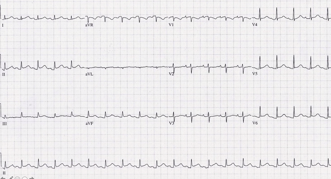
An ecg is done and is shown below.
​What is the diagnosis?
Picture
This is atrial flutter. It is the second most common tachy-arrhythmia after atrial fibrillation
It is a macroreentrant atrial tachycardia with the loop above the AV node in the right atrium.
  • type 1 flutter is the most frequent.
    • In this type of flutter, right atrial activation rotates in a counterclockwise direction. 
It usually produces a narrow complex tachycardia, with a classic sawtooth pattern best seen in lead II.
It can conduct as
  • 1:1, which can be unstable
  • 2:1, which is the most common
  • 3:1
  • 4:1
  • It can also be variable which gives the appearance of an irregular rhythm.
How do we best manage the STABLE patient?
How to best manage the UNSTABLE patient?
​Who would you anticoagulate?
Remember that this arrhythmia will typically not revert with vagal manoeuvres.
MEMBERS CLICK HERE FOR MORE
0 Comments

A 68 YO WOMAN WITH WORSENING SOB

15/6/2021

0 Comments

 
This year at the Cardiac Bootcamp face to face course in Sydney we had a fantastic turnout and we went over a huge amount of content in Acute Cardiology and reading ECGs.
There were some ECGs that we looked at, as part of the ECG Quiz.
​Here is one of those ECGs, to test your memory and to reinforce some of the most important things we learnt about this case.
A 68 yo woman presented with worsening SOB over the previous 24 Hours. She now also had chest pain and was speaking in very short phrases.
​She was a smoker, with a past medical history of COPD and hypertension.
Her vitals were:
  • HR 115
  • BP 87/42
  • RR 31
  • Sats 94% on room air
  • Temp 36.2
Her examination showed dual heart sounds, with some crepitations in the right base and her abdo was soft.
Her blood pressure dropped to 65/32 before peripheral inotropes were commenced.
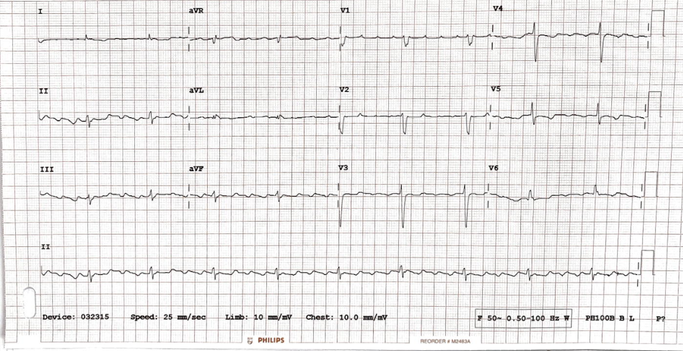
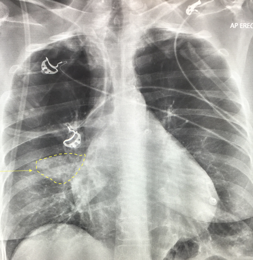
Her ECG and Chest Xray are shown below. What is your diagnosis?
Picture
Picture
There is a pericardia effusion diagnosed on LOW VOLTAGE CRITERIA.
Small complexes and tachycardia = pericardial effusion.
The are Sensitive Criteria and Specific criteria for picking low voltage on an ECG.
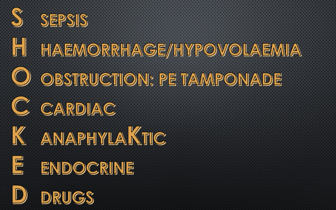
This was all part of the 'SHOCKED' A mnemonic on Cardiac Bootcamp Course, to minimise missed cases.
Picture
MEMBERS CLICK HERE FOR MORE
0 Comments

A 50 yo with chest pain

8/6/2021

0 Comments

 
A 50 yo male presents with chest pain. His ECG is shown below. What does it show?
Picture
Rate: 66
Rhythm: Irregularly irregular, not many P waves, so atrial fibrillation.
QRS: Narrow
ST-T: The obvious abnormality here is the STE in II, III and aVF with reciprocal changes in aVL.
There is also some ST depression in V2-V4.

The diagnosis here is an inferior infarction. We need to beware of potential Right Ventricular infarction as well.
How do patients with Right Ventricular Infarction present?
What does the ECG show?
What are the differentials?
What is the management of these patients?
​
Isolated right ventricular infarction is rare, but it does occur in 30-50% of cases of inferior infarction. We need to think of the possibility of right ventricular infarction, or we can miss it.
The right ventricle is thin walled and is perfused throughout the cardiac cycle, explaining why it is less likely to infarct.......
CARDIAC BOOTCAMP MEMBERS READ MORE
0 Comments

    Author

    Dr Peter Kas
    ​Emergency Physician

    Picture
    Picture
    Sign up for
    ECG of the week
     Its free

    Archives

    February 2023
    January 2023
    December 2022
    November 2022
    October 2022
    September 2022
    August 2022
    July 2022
    June 2022
    March 2022
    December 2021
    November 2021
    October 2021
    September 2021
    August 2021
    July 2021
    June 2021
    May 2021
    April 2021
    March 2021
    February 2021
    January 2021
    December 2020
    November 2020
    September 2020
    August 2020
    July 2020

    Categories

    All
    Atrial Fibrillation
    Atrial Flutter
    Atrial Tachycardia
    Axis
    Bradycardia
    Cardiac Arrest
    Chest Pain
    ECG Mimics
    Ischaemia
    Lead Reversal
    Myopericarditis
    Pacing
    Paediatric Arrhythmias
    Paediatric ECG
    Paediatric Syncope
    Pericarditis
    QT Interval
    STEMI
    STEMI Mimics
    SVT
    Ventricular Tachycardia
    Wellens
    Wide Complex Tachycardia

Our Other Websites to Visit

www.emcore.com.au
www.resus.com.au

Contact Us
© COPYRIGHT 2015. ALL RIGHTS RESERVED.
  • HOME
  • Course Login
    • Log In
    • Log Out
  • SYLLABUS
  • STORE
    • Online Self Study Course
    • Cardiac Video Course
  • ECG OF THE WEEK