CARDIAC BOOTCAMP
  • HOME
  • Course Login
    • Log In
    • Log Out
  • SYLLABUS
  • STORE
    • Online Self Study Course
    • Cardiac Video Course
  • ECG OF THE WEEK

ECG OF THE WEEK

BECOME A COURSE MEMBER
Each Week we put up one ECG case for you....because it's easier to learn from cases.
The ECG of the week is FREE to everyone. To become an expert at ECG's join the Cardiac Bootcamp Course. It's a great way to Learn!
​Or come to one of our LIVE Cardiac Bootcamps

cardiac arrest pearls

30/8/2021

0 Comments

 
A 68 year old male is brought into resus with chest pain and diaphoresis. The patient rapidly loses consciousness and is in cardiac arrest, with the rhythm strip shown below. CPR was rapidly commenced and the team went into resus mode.
Picture
This week we look at a few pearls that come straight out of the 'Cardiac Arrest Pearls' part of the Cardiac Bootcamp Course. Some of these may surprise you.....
​

The Pulse Check
During the rhythm check during CPR someone checked the pulse.

During the rhythm check someone checked the pulse.
Can we rely on the pulse to guide further resuscitative efforts?
We must stop relying on the pulse.
Old ATLS teaching was it that a carotid or femoral pulse being present, meant that the systolic blood pressure was at least 70mmHg.
Studies have shown that our ability to detect a pulse and the accuracy of the reading are unreliable. Also the blood pressure with a pulse present can be less than 60mmHg
Deakin et al BMJ 2000 Sept 16;321(7262):673-674
CPR
CPR is everything and distinguishes between NO FLOW(no CPR) and LOW FLOW(CPR). At best we will achieve 50% of normal cardiac output (yes, that's with the best CPR we can perform). It's not great, but it again reinforces the need to continue CPR and not interrupt it.
The QUALITY OF CPR IS DEFINED BY:
  1. Chest compression FRACTION
  2. Chest compression RATE
    1. A quick word on RATE OF CPR:
      100 compressions per minute is the recommended rate, however data from in-hospital cardiac arrests have shown that when the rate is too slow, return of spontaneous circulation(ROSC) falls from 72% to 42%
      Animal models have shown that faster rates lead to better cardiac output.
      A 2017 prospective observational study that measured ROSC in in-hospital cardiac arrests found that the greatest chance of ROSC was with a rate of 121-140. Resuscitation Jan 2017 Vol 110 pp154-161
  3. Chest compression DEPTH
  4. Chest RECOIL
Go to the Cardiac Arrest Pearls Section to find out more.

AIRWAY
Everyone is obsessed with "getting the tube in". In resuscitations, the airway is simple, unless there are compelling reasons to; do not intubate until ROSC is achieved. Intubation confers no benefit over supraglottic airway.
SOME POINTS ON AIRWAY
  • We don't know the best time to intubate
  • It is difficult to intubate without interrupting CPR. This is an important point. If you have a video laryngoscope and can coordinate your intubation with CPR, then go ahead. However do NOT interrupt CPR to intubate
  • Without adequate ETCO2, as occurs during cardiac arrest, it is difficult to very Ett placement.
  • I BVM the patient and intubate only when ROSC is achieved.

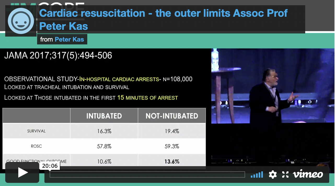
All Patients should be intubated as soon as possible following a cardiac arrest: True/False?
ANSWER
FALSE
The studies in out of hospital cardiac arrest showed that intubation conferred no benefit over BVM or supraglottic airway. In in-hospital cardiac arrests, those patients intubated within the first 15 minutes of cardiac arrest, had a worst outcome.
ADRENALINE
The use of 1 mg of adrenaline which initially came from canine models, was introduced at this time and has been a major part of all resuscitations since then.
The clinical significance of adrenaline is uncertain. It’s been shown to give no improvement and even to decrease survival to hospital discharge and decrease the rate of favourable neurological outcomes. Steil in the multi-centre OPALS study demonstrated no improvement in neurologically intact survivors to hospital discharge when using advanced life-support. We know that high-dose adrenaline doesn’t improve outcomes.
Jacobs, further randomised 601 cardiac arrest patients and found a significantly improved likelihood of achieving return of spontaneous circulation(ROSC) in the adrenaline group. There was a trend to increased survival to discharge in this group, unfortunately the study was underpowered.

Everyone should get 1mg of Adrenaline right?
Giving 1mg of adrenaline to a heart that has just resumed beating, can have dire consequences. The effect of markedly increased afterload against which a weak heart must beat, can result in a rapid loss of circulation. The adrenaline dose must be titrated to achieve adequate coronary and cerebral perfusion.
Our aim should be to achieve a coronary perfusion pressure of above 15 – 20 mmHg. This equates to diastolic blood pressure of 25 – 35 mmHg, which has been shown to provide adequate cerebral perfusion pressure.
Monitoring with an arterial line is the only way to do this accurately. As I have discussed at several EMCORE conferences, the aim is to locate the femoral artery with ultrasound during the first rhythm check pause and to then pass the catheter during the second rhythm check. This requires practice and coordination of the team.
If you don’t have an arterial line, don’t trust the pulse check, or the electrical wave form. Use the cardiac ultrasound to verify whether the heart is beating or not.
In the FEEL study it was found the 38% of patients with asystole on the ECG, had coordinated cardiac motion on echo. It was further found that 58% of those in PEA had coordinated cardiac motion seen when the heart was visualised. Hypothesize what might happen with the massive alpha effects of giving 1 mg of adrenaline to these hearts. Keep the dose small if the heart is beating.
Cardiac Bootcamp Members go to the Cardiac Pearls Section for More PLUS watch the Video
Picture
JOIN THE CARDIAC BOOTCAMP COURSE
0 Comments

WHAT IF WE COULD PREDICT BY EVOLVING ECG CHANGES WHO WAS GOING TO ARREST?

6/7/2021

0 Comments

 
What if we had a way to predict PEA/Asystolic Cardiac Arrest, a few minutes before it happened? Would that be helpful?.... Perhaps.
PEA and asystolic cardiac arrests have a very poor outcome. The rate of survival in out of hospital arrests is less than 2%. In hospital, survival is higher, at about 30%(1).
Having this knowledge may or may not change the outcome, but I think it gives us a significant advantage, at least in our mental preparation.

In a recent research paper by Do et al in Resuscitation(2), they identified a right ventricular strain pattern that preceded hypoxic pulseless electrical activity cardiac arrest.

STUDY DESIGN: What they did.
This was a retrospective cross sectional study at two tertiary care hospitals. They looked at in hospital cardiac arrests due to PEA/Asystole
  • Patients were at least 18 years of age
  • There was telemetry data for at least 3 hours prior to the arrest.
  • n=140
Patients were excluded if they had:
  • pulmonary hypertension
  • right ventricular enlargement on ECG,
  • ventricular paced rhythm,
  • an out of hospital arrest leading to the current admission,
  • this was not their first in hospital cardiac arrest.

Right Ventricular strain pattern
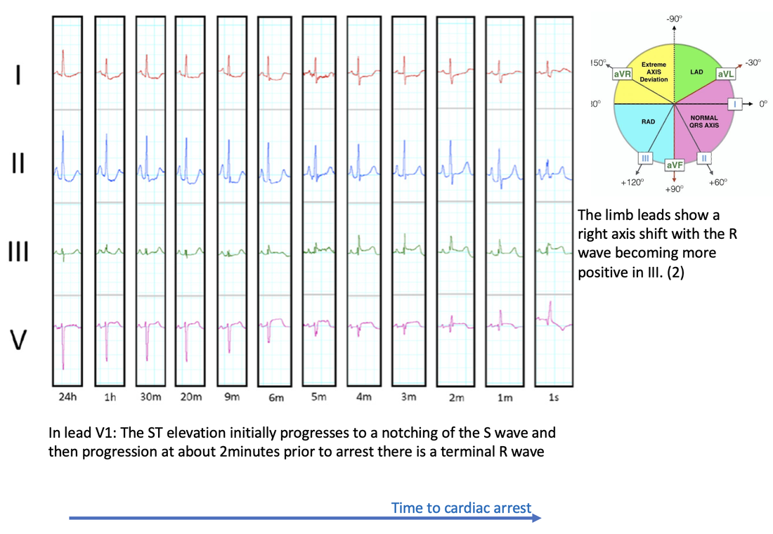
The most common terminal morphology(67% of cases) was a RBBB in V1, which progressed from an increasing RV delay as shown below:
Picture
Definite Right Ventricular strain(RVS) pattern was defined as having the above progressive morphological changes in lead V1 and either no intraventricular conduction delay or terminal intraventricular conduction delay (ie., incomplete RBBB) and two or more of the following occurring simultaneously:
  1. ST elevation V1
  2. Rightward directed ST elevation vector in limb leads (i.e. towards lead III) or
  3. Right axis deviation in limb leads
Possible RVS was defined as the RV delay pattern in V1 and only one of the supporting features.

Results
  • The median time of RVS pattern to appear on the ECG in those that had a previously normal baseline was 7.2 minutes before arrest.
  • The RVS pattern prior to PEA/Asystolic arrest was present in 47% of patients, without pre-existing right ventricular hypertrophy
  • In those patients with RVS pattern who went on to have a cardiac arrest and then ROSC, there was a 41% incidence of RV dysfunction on echo.
​
ECG Patterns identified
Picture
Conclusion
Next time we are monitoring a patient and see the gradual Right ventricular delay pattern, indicative of right ventricular failure, we should get ready as up to about half of these patients may have a PEA/Asystolic arrest in the next few minutes.

REFERENCES
  1. Holmberg Mathias J et al. Annual incidence of adult and paediatric in-hospital cardiac arrest in the United States. Circ Cardiovasc Qual Outcomes 2019; 12:e005580.
  2. Do D H. et al. Electrocardiographic right ventricular strain precedes hypoxic pulseless electrical activity cardiac arrests; Looking beyond pulmonary embolism. Resuscitation151(2020) pp 127-134.
0 Comments

    Author

    Dr Peter Kas
    ​Emergency Physician

    Picture
    Picture
    Sign up for
    ECG of the week
     Its free

    Archives

    February 2023
    January 2023
    December 2022
    November 2022
    October 2022
    September 2022
    August 2022
    July 2022
    June 2022
    March 2022
    December 2021
    November 2021
    October 2021
    September 2021
    August 2021
    July 2021
    June 2021
    May 2021
    April 2021
    March 2021
    February 2021
    January 2021
    December 2020
    November 2020
    September 2020
    August 2020
    July 2020

    Categories

    All
    Atrial Fibrillation
    Atrial Flutter
    Atrial Tachycardia
    Axis
    Bradycardia
    Cardiac Arrest
    Chest Pain
    ECG Mimics
    Ischaemia
    Lead Reversal
    Myopericarditis
    Pacing
    Paediatric Arrhythmias
    Paediatric ECG
    Paediatric Syncope
    Pericarditis
    QT Interval
    STEMI
    STEMI Mimics
    SVT
    Ventricular Tachycardia
    Wellens
    Wide Complex Tachycardia

Our Other Websites to Visit

www.emcore.com.au
www.resus.com.au

Contact Us
© COPYRIGHT 2015. ALL RIGHTS RESERVED.
  • HOME
  • Course Login
    • Log In
    • Log Out
  • SYLLABUS
  • STORE
    • Online Self Study Course
    • Cardiac Video Course
  • ECG OF THE WEEK