CARDIAC BOOTCAMP
  • HOME
  • Course Login
    • Log In
    • Log Out
  • SYLLABUS
  • STORE
    • Online Self Study Course
    • Cardiac Video Course
  • ECG OF THE WEEK

ECG OF THE WEEK

BECOME A COURSE MEMBER
Each Week we put up one ECG case for you....because it's easier to learn from cases.
The ECG of the week is FREE to everyone. To become an expert at ECG's join the Cardiac Bootcamp Course. It's a great way to Learn!
​Or come to one of our LIVE Cardiac Bootcamps

Diagnosing Left Ventricular hypertrophy: The new seamens' sign

27/7/2022

0 Comments

 
We know that Left Ventricular Hypertrophy (LVH) when diagnosed on an ECG, is associated with a higher mortality and cardiovascular morbidity.
The gold standard for diagnosis is Cardiac Echocardiography, or ventricular mass measurements by MRI.
The ECG however can provide a helpful screening tool for patients, alerting us for the need for further investigations.

How do you determine if the ECG shows LVH?
MEMBERS  go to LVH under the ECG section to read about:
  • CORNELL CRITERIA
  • SOKOLOW-LYON CRITERIA
  • ROMHILT-ESTES POINT SCORE SYSTEM
A new study in Peer Journal, looked at the new Seamens' Criteria.
​
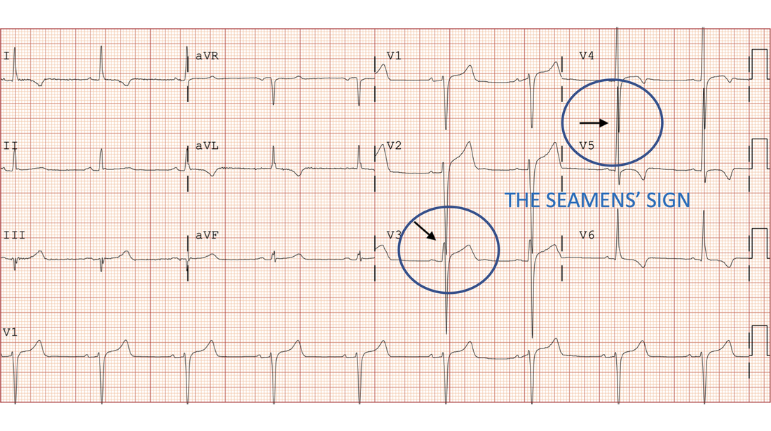
The SEAMENS' SIGN
This is a new proposed sign from the recent literature(1).
The Seamens Sign is as follows:
If any of the QRS Complexes in the precordial leads in a 12 lead ECG, touch or cross another QRS complex, this is defined as LVH.

​
This was a retrospective chart review of 2184 patient records in a Quartenary Centre and looked at consecutive patients with an ECG and a trans thoracic echocardiogram, performed within 90 days of each other.
The Primary Endpoint was to determine if the Seamens' Sign was non inferior in confirming LVH compared to other criteria.
Conclusion
When compared to the Sokolow-Lyon criteria and the Cornell criteria for males, the Seamens' Sign was non-inferior in confirming LVH on an ECG. Below is the example.
Picture
0 Comments

A 12 yo with collapse.

20/7/2022

0 Comments

 
Syncope is not uncommon, occurring in up to 25% of children before 18 years of age. Most causes are benign, however we need to be aware of the of 3 major groups as these can be very serious diagnoses:
  1. Those with a cardiac cause
  2. Those with a neurological cause
  3. Very young children, as syncope shouldn’t occur in this group.
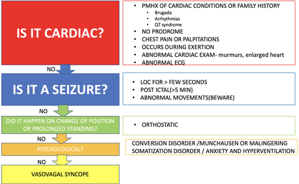
Making the diagnosis in children can be stressful, so I like to use a simple algorithmic approach to work through the possible diagnoses. This is primarily so I don’t miss the more important causes. There are syncope rules in existence, however these apply to adult patients.

Definition of Syncope
​
Syncope in children and adults is a symptom, not a condition. It is up to us to determine the cause. The definition of syncope involves three components:
  1. Loss of consciousness
  2. Loss of postural tone
  3. Recovery and return to baseline
This third part of the definition is important. Simple neurocardiogenic syncope will have a return to baseline usually within about 30 seconds. However if there is altered consciousness for more than about 5 minutes, then seizure or other neurological cause needs to be considered.

Case

A 12 yo presents to the emergency department. He is unwell and has had some episodes of syncope. His investigations in the ED result in the diagnosis of Viral Encephalitis.

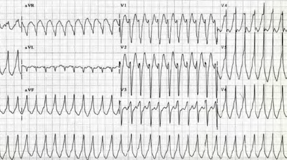
He develops the rhythm shown below and at the same time drops his conscious state. What does the ECG show? What is the diagnosis? What would you do now?
Picture
What is the diagnosis?
Ventricular Tachycardia

Do you have an approach to Paediatric Syncope?

Members login and go to the Syncope in Children section.
Below you will find my approach to the child with syncope. Read more on this on the website, and also watch the video lecture.
Picture

Watch the Lecture

Picture
0 Comments

A 40 yo in cardiac arrest

9/7/2022

0 Comments

 
A 40 yo male is brought in by ambulance following a collapse
His GCS at the scene was 3
Paramedics found his rhythm to be VT? and he was given 11 shocks and 5mg of Adrenaline in total.
On arrival he has an undetectable BP. He is maxed out on an adrenaline infusion.
He has an LMA and has spontaneous assisted respirations
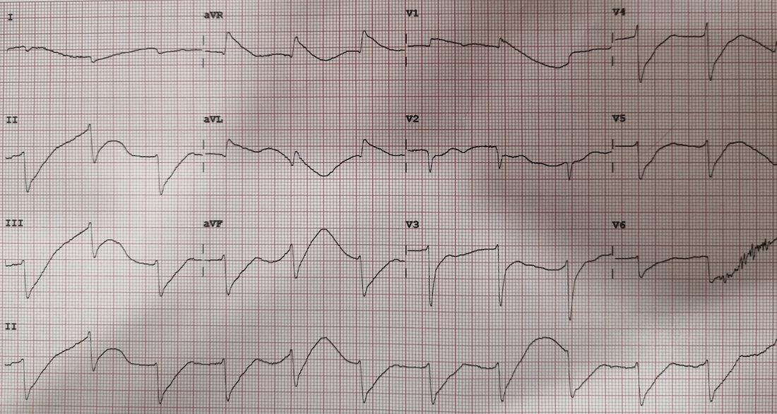
The ECG is shown below. What do you think are the important changes
Picture
What would you treat this patient with, acutely? He is maxed out on an adrenaline infusion.
(a)Adrenaline
(b)
Calcium Gluconate
(c)Amiodarone
(d)Potassium
(e)Atropine

ANSWER
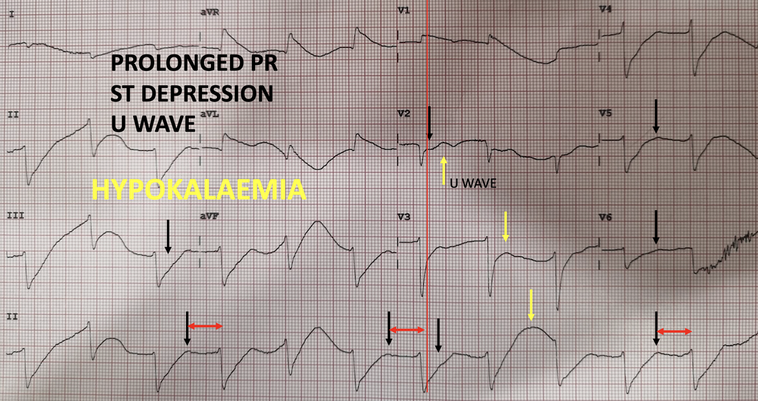
The answer is d. This is the ECG of hypokalaemia.
Notice the PR prolongation, ST depression and U waves.
See the ECG changes below.
.
.
.
.
​.
Picture

What are the causes of this condition?
What is the treatment?
Was it really VT or something else?
​What is the issue with Adrenaline in this case?
Cardiac Bootcamp Members go to the Hypokalaemia section for answers and for another case.
0 Comments

A 38 yo male presents with chest pain

2/7/2022

1 Comment

 
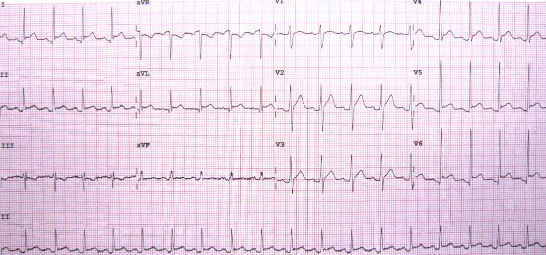
A 38 yo male presents with sharp stabbing central chest pain. It changes with breathing. He feels worst when he is lying down. He is haemodynamically stable. An ECG is done. What does it show?
Picture
WHAT DOES THE ECG SHOW?
Using the ECG in 20 Seconds Approach:
  • There is a sinus tachycardia at about 108bpm
    • The P wave is upright in II and inverted in aVR, so it is sinus
  • The QRS morphology is normal
  • The ST-T segments are not normal:
    • There is ST elevation in multiple leads ie., I, II aVL, V4, V5, V6
    • There appears to be a J point in aVL, V4, V5 and V6, so Benign early repolarisation is an alternative diagnosis.
    • There is ST depression in aVR
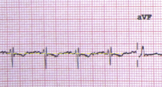
    • Is that ST depression in III?​
      • Initially it does look like ST depression, however when we draw in the isoelectric line, it is not really as significant (see below).
Picture
REMEMBER: When looking for ST elevation or depression, use the T-P line as the isoelectric line.
  • The QT and PR intervals are normal
    • It is obvious that there is PR depression
  • There are no pacing spikes
DIAGNOSIS
The diagnosis is most likely pericarditis.
The history is very indicative of that. The characteristics of the pain ie., that is it worst on lying down indicate pericarditis. Although there appears to be a pleuritic component, there is no real other history of a PE and the ECG does not show a right axis or S1Q3T3 etc.

In the ECG itself:
  • There is ST elevation, concave in nature
  • There is ST depression in aVR, which is allowed ie., there can be ST depression in aVR and V1
  • There are no ischaemic looking waveforms and no real reciprocal changes.
  • There is PR depression.
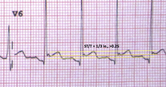
  • We can also measure the ST/T ratio. A ratio of >0.25, when put together with all the other parameters indicates pericarditis.
Picture
WHAT TO DO NEXT
  • Do a bedside echo looking for an effusion. It will also assist to look for wall abnormalities.
  • Get a troponin. A slight rise in troponin may be normal. We need to make sure that we don't miss, perimyocarditis or myocarditis, where we would expect a significant rise in troponin.
Who needs admission and who can be discharged?
What is Spodick's Sign and how good is it?
What are the stages of Pericarditis?
What is the treatment?
​Is there a role for glucocorticoids?
Members login and go to the pericarditis section for answers to all the above and more
1 Comment

    Author

    Dr Peter Kas
    ​Emergency Physician

    Picture
    Picture
    Sign up for
    ECG of the week
     Its free

    Archives

    February 2023
    January 2023
    December 2022
    November 2022
    October 2022
    September 2022
    August 2022
    July 2022
    June 2022
    March 2022
    December 2021
    November 2021
    October 2021
    September 2021
    August 2021
    July 2021
    June 2021
    May 2021
    April 2021
    March 2021
    February 2021
    January 2021
    December 2020
    November 2020
    September 2020
    August 2020
    July 2020

    Categories

    All
    Atrial Fibrillation
    Atrial Flutter
    Atrial Tachycardia
    Axis
    Bradycardia
    Cardiac Arrest
    Chest Pain
    ECG Mimics
    Ischaemia
    Lead Reversal
    Myopericarditis
    Pacing
    Paediatric Arrhythmias
    Paediatric ECG
    Paediatric Syncope
    Pericarditis
    QT Interval
    STEMI
    STEMI Mimics
    SVT
    Ventricular Tachycardia
    Wellens
    Wide Complex Tachycardia

Our Other Websites to Visit

www.emcore.com.au
www.resus.com.au

Contact Us
© COPYRIGHT 2015. ALL RIGHTS RESERVED.
  • HOME
  • Course Login
    • Log In
    • Log Out
  • SYLLABUS
  • STORE
    • Online Self Study Course
    • Cardiac Video Course
  • ECG OF THE WEEK