CARDIAC BOOTCAMP
  • HOME
  • Course Login
    • Log In
    • Log Out
  • SYLLABUS
  • STORE
    • Online Self Study Course
    • Cardiac Video Course
  • ECG OF THE WEEK

ECG OF THE WEEK

BECOME A COURSE MEMBER
Each Week we put up one ECG case for you....because it's easier to learn from cases.
The ECG of the week is FREE to everyone. To become an expert at ECG's join the Cardiac Bootcamp Course. It's a great way to Learn!
​Or come to one of our LIVE Cardiac Bootcamps

A 60 yo male collapses at work

22/10/2021

0 Comments

 
A 60 yo male is brought into the Emergency Department following collapsing at work.
On arrival he is alert and oriented with a heart rate of 20bpm, BP of 105/70 and a respiratory rate of 16.
He has no chest pain or other concerning features.
His past history includes:
  • Atrial Fibrillation (on Sotalol and a NOAC)
  • Hypercholesterolaemia
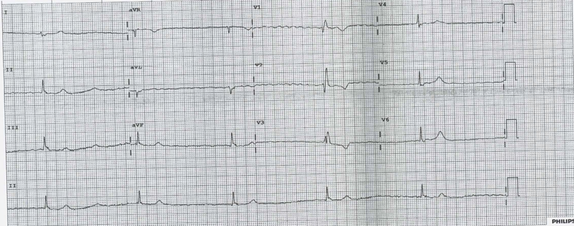
His ECG is shown below. After looking at his ECG, what would you do?
​

(a) Increase his heart rate using pharmacology
(b) Do nothing as you might cause harm and his BP is acceptable and he is alert.
(c) Defibrillate
(d) Give Magnesium or an extra dose of Sotalol
Picture
The ECG shows a slow regular (junctional) rhythm with narrow complexes and a RBBB. 
Beware assuming that this patient's cardiac output is fine, given that he is alert and his blood pressure is 'acceptable'. The cardiac output MUST be reduced. CO = HR x SV  The only reason the blood pressure is not low is due to a sympathetic response mounted by the patients, resulting in increased vasoconstriction. 

(a) Increase his heart rate using pharmacology- The only real answer
(b) Do nothing as you might cause harm and his BP is acceptable and he is alert. X the patients cardiac output is reduced and they are only perfusing due to endogenous sympathetic response.
(c) Defibrillate X
(d) Give Magnesium or an extra dose of Sotalol X
​

Pharmacological Increases in Heart Rate

 ATROPINE
Muscurinic Acetylcholine Receptor Antagonist
t1/2 = 3-4 hours
Indication: AV nodal blockade. It works by decreasing vagal tone, so may not be effective in high degree AVN block.
Dose: Start at 0.5 mg-1mg (max of 3mg) Lower doses may cause a reflex bradycardia
Beware:
  •  Patients with coronary artery disease as can increase myocardial demand​
  • Acute angle closure glaucoma
  • Bowel obstruction
  • Cardiac transplant patients- may result in asystole.
Atropine may be the first line to use whilst you are getting set up for the definitive drug ie Isoprenaline. Atopine may not work on all patients or if it does work, beware, as it can wear off fairly rapidly.

ISOPRENALINE
Beta adrenoreceptor agonist 
t1/2 = 5 minutes, but immediate onset of action
Dose: If given centrally start at 0.5-1 mcg/min and titrate. It is also safe for peripheral infusion and can be started at 5mcg/min then titrate up or down. Usual range is 2-10 mag/min, however higher doses have been used.
Beware: Hypotension, tachycardia, angina. May not be tolerated by the patient longer term as can cause headaches and trembling.

ADRENALINE
​Non-selective adrenergic agonist Beta 1 > alpha 1 and Beta 2
t1/2 = 5 minutes. Acts within a minute and lasts for 5-10 minutes.
Dose: This can be used peripherally. Start dose at 1 mcg/min. Usual dose is 1-10 mcg/min. Higher doses may be needed.
It has a wide range of effect, being a chronotrope and inotrope.
Beware: 
​Tachycardia and arrhythmias, narrow angle glaucoma, Phaeochromocytoma

​.........MEMBERS can go to the new section titled Managing Bradycardia and read further about Transcutaneous and Transvenous Pacing
Members can also read about Blocks

What happened to the patient?

He responded well to Isoprenaline..perhaps too well, becoming a little tachycardia. However given that isoprenaline has such a rapid turnoff effect, it was weaned. The patient was discussed with cardiology and was referred for a pacemaker.
Picture
0 Comments

What is pseudo-wellens' Syndrome?

13/10/2021

1 Comment

 
Wellen's Syndrome involves an ECG pattern representing a significant major coronary artery stenosis. More specifically, a Left Anterior Descending(LAD) artery stenosis. In most cases this is proximal, but it may be mid or distal LAD.
It actually represents a reperfusion of the vessel and is one more reason to perform serial and painfree ecgs. If not recognised and treated, it  can result in up to 75% of patients having an anterior wall myocardial infarction within days/weeks. 


How to Recognise the Pattern?
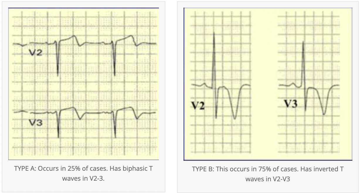
The peculiarity of this ECG pattern is that it may not appear when the patient has pain. In fact the only finding when the patient has chest pain, may be a small negative deflection at the end of V1 and V2 . The T waves become deeply inverted when the patient is pain free and represents a reperfusion of the myocardium. Cardiac enzymes will be normal in most cases or mildly elevated.
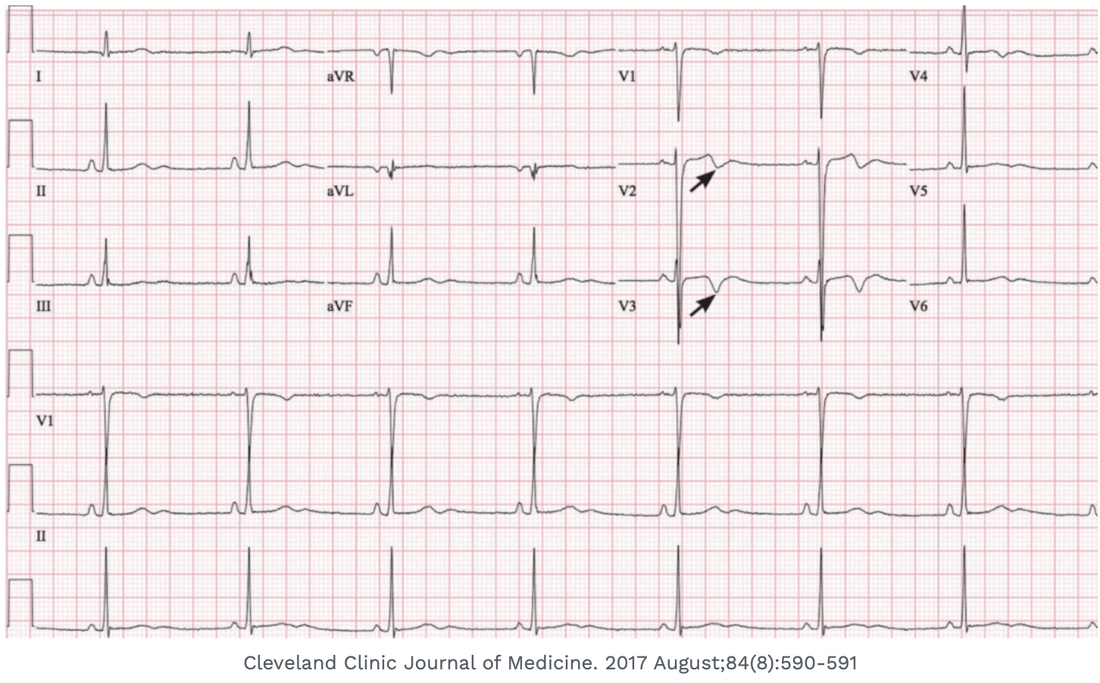
Below are the two key ecg patterns in Wellens'.
Picture

What is Pseudo-Wellens'?

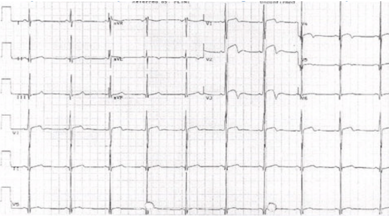
It is a mimic. Pseudo-Wellens’, is a similar pattern to Wellens’, but results from other ECG abnormalities such as  abnormal T waves, due to left ventricular hypertrophy(LVH). We cannot diagnose Wellens' Syndrome in a patient with left ventricular hypertrophy. The biphasic waveforms are a direct result of the left ventricular hypertrophy, and represent a T wave change associated with the enlarged heart.
So if there is LVH we cannot diagnose Wellens’ Syndrome on ECG. The patient may however still have ischaemia, so beware. One more thing that is different in Pseudo-Wellens’, is that the changes are more lateral i.e.., V3-V6, whereas in true Wellens’ they are at V2-V3.
The ECG below is a very good example of the ‘pseudo’ pattern.  The biphasic waveforms are due to left ventricular hypertrophy. They are also more lateral than the true Wellens. 
Picture

Other mimics also exist

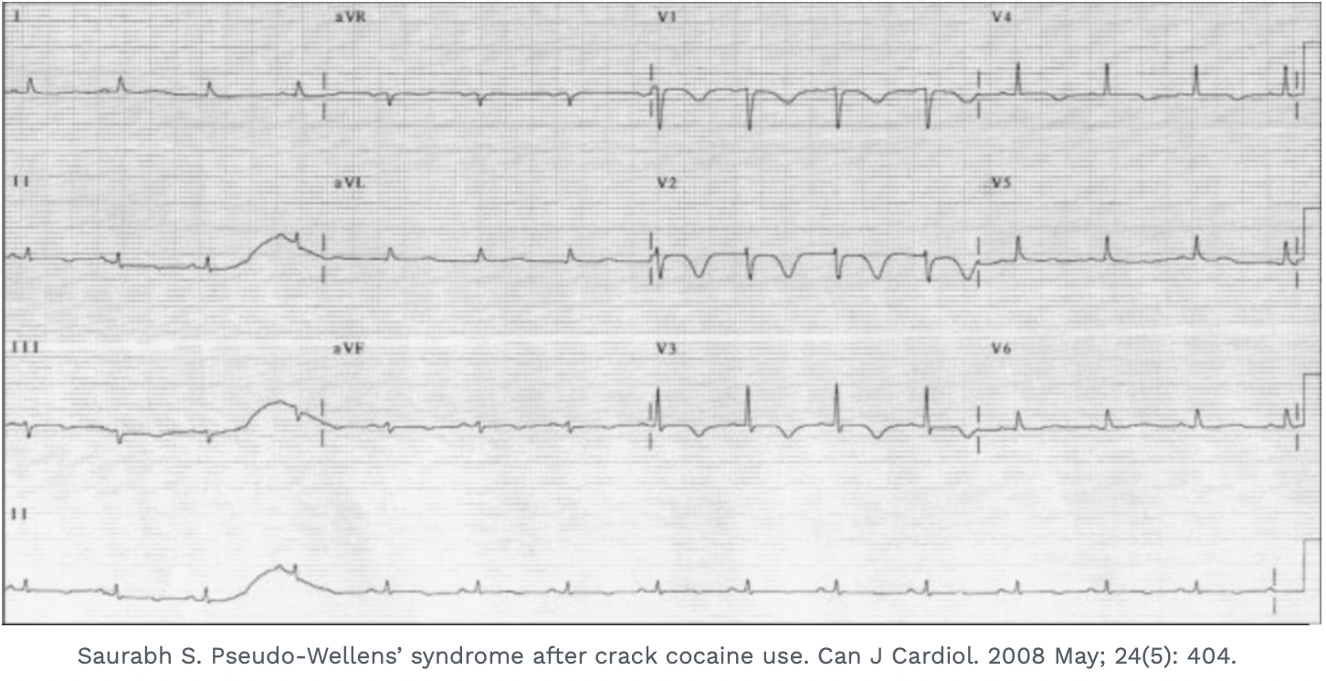
Substance abuse such as cocaine and other stimulants as well as cannabis can also result in a mimic pattern. Examples of these mimics are shown below.
Even though we may consider them as mimics, our first priority always is to rule out ischaemia first. The diagnosis of a pseudo-Wellens' is a diagnosis of exclusion.
A patient presenting after taking cocaine and showing the pseudo-Wellens' pattern:
Picture
A patient with excessive cannabis use, showing a pseudo-Wellens' pattern:
Picture
1 Comment

That's a little fast isn't it?!!!

8/10/2021

0 Comments

 
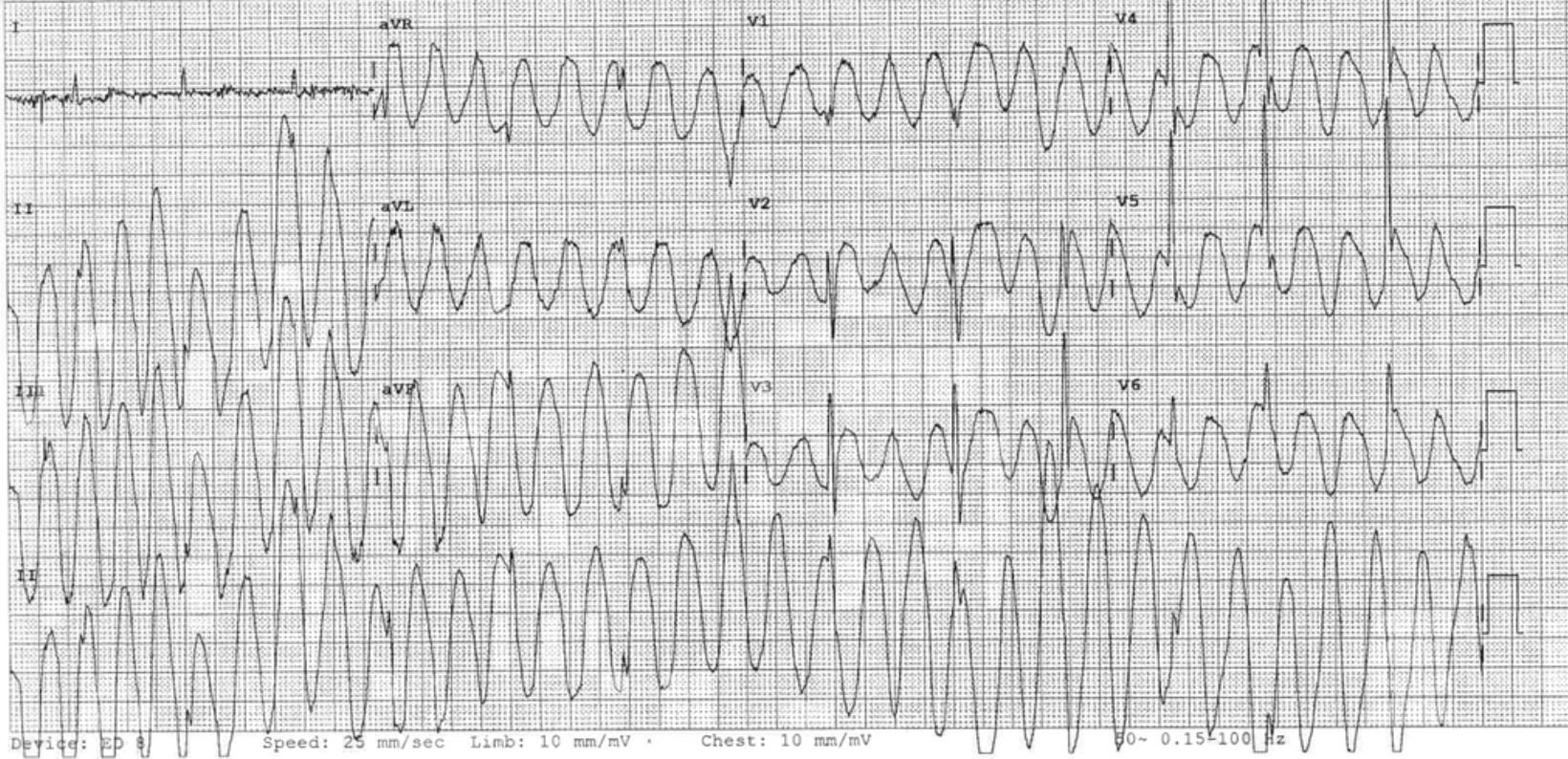
A 90 yo patient with multiple co-morbidities and mild dementia presents with chest pain.
The patient is haemodynamically stable. 
An ECG is done and there is concern over the rate. 
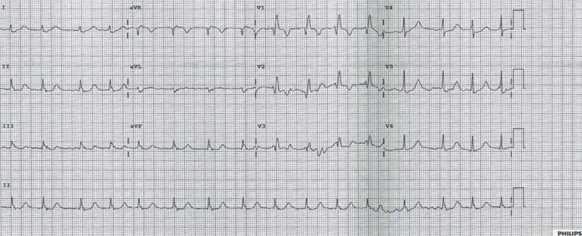
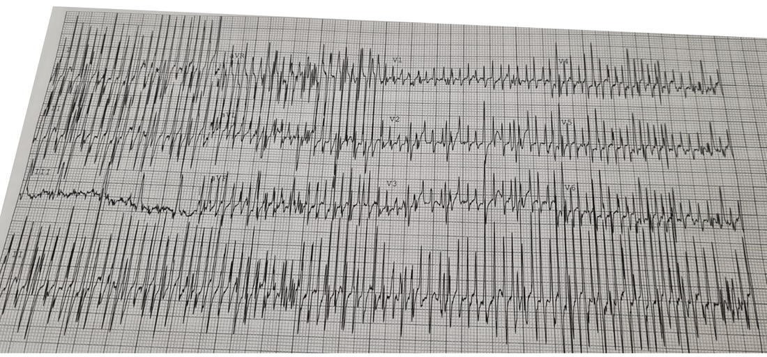
Look at the ECG. What is the diagnosis?
Picture
Is it SVT? Is it Torsades? What do you do next?
.........Move away from the patient..... ie., do nothing. Let's see why.
Let's go back to the clinical picture.
The patient is haemodynamically stable and looks well.
Now back to the ECG: It is a narrow complex EXTREME tachycardia.
Notice lead III. It is not the same as the others. One lead cannot refuse to show the tachycardia whilst all the others are. In fact lead III here is the underlying rhythm. The rest of the ECG is artifact.
Remember that if the ventricle is beating at above 300 beats per minute in an adult patient, it will very soon convert to ventricular fibrillation.
Clinically at a rate of 350 or so, the patient will not be stable, as the ventricle doesn't have time to fill properly, so the blood pressure will be impaired. 

What is the artifact caused by?
Artifacts can be external, relating to line current. They can also be internal relating to tremor or shivering. They can also be caused by other devices such as deep brain stimulators ( which are usually there to control tremor).

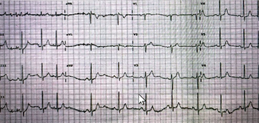
​An ECG was redone and is shown below. The artifact is gone and the patients underlying rhythm is there.
Picture
Don't be fooled by the mimics.
To read more on this and see more examples like the one below go to the blog on 'A Patient with Wide Complex Tachycardia'
Picture
The Face to Face Cardiac Bootcamp is on the Gold Coast in 2022
0 Comments

    Author

    Dr Peter Kas
    ​Emergency Physician

    Picture
    Sign up for
    ECG of the week
     Its free

    Archives

    December 2022
    November 2022
    October 2022
    September 2022
    August 2022
    July 2022
    June 2022
    March 2022
    December 2021
    November 2021
    October 2021
    September 2021
    August 2021
    July 2021
    June 2021
    May 2021
    April 2021
    March 2021
    February 2021
    January 2021
    December 2020
    November 2020
    September 2020
    August 2020
    July 2020

    Categories

    All
    Atrial Fibrillation
    Atrial Flutter
    Atrial Tachycardia
    Axis
    Bradycardia
    Cardiac Arrest
    Chest Pain
    ECG Mimics
    Ischaemia
    Lead Reversal
    Pacing
    Paediatric ECG
    Paediatric Syncope
    Pericarditis
    QT Interval
    STEMI
    STEMI Mimics
    SVT
    Ventricular Tachycardia
    Wellens
    Wide Complex Tachycardia

Our Other Websites to Visit

www.emcore.com.au
www.resus.com.au

Contact Us
© COPYRIGHT 2015. ALL RIGHTS RESERVED.
  • HOME
  • Course Login
    • Log In
    • Log Out
  • SYLLABUS
  • STORE
    • Online Self Study Course
    • Cardiac Video Course
  • ECG OF THE WEEK