CARDIAC BOOTCAMP
  • HOME
  • Course Login
    • Log In
    • Log Out
  • SYLLABUS
  • STORE
    • Online Self Study Course
    • Cardiac Video Course
  • ECG OF THE WEEK

ECG OF THE WEEK

BECOME A COURSE MEMBER
Each Week we put up one ECG case for you....because it's easier to learn from cases.
The ECG of the week is FREE to everyone. To become an expert at ECG's join the Cardiac Bootcamp Course. It's a great way to Learn!
​Or come to one of our LIVE Cardiac Bootcamps

cardiac arrest pearls

30/8/2021

0 Comments

 
A 68 year old male is brought into resus with chest pain and diaphoresis. The patient rapidly loses consciousness and is in cardiac arrest, with the rhythm strip shown below. CPR was rapidly commenced and the team went into resus mode.
Picture
This week we look at a few pearls that come straight out of the 'Cardiac Arrest Pearls' part of the Cardiac Bootcamp Course. Some of these may surprise you.....
​

The Pulse Check
During the rhythm check during CPR someone checked the pulse.

During the rhythm check someone checked the pulse.
Can we rely on the pulse to guide further resuscitative efforts?
We must stop relying on the pulse.
Old ATLS teaching was it that a carotid or femoral pulse being present, meant that the systolic blood pressure was at least 70mmHg.
Studies have shown that our ability to detect a pulse and the accuracy of the reading are unreliable. Also the blood pressure with a pulse present can be less than 60mmHg
Deakin et al BMJ 2000 Sept 16;321(7262):673-674
CPR
CPR is everything and distinguishes between NO FLOW(no CPR) and LOW FLOW(CPR). At best we will achieve 50% of normal cardiac output (yes, that's with the best CPR we can perform). It's not great, but it again reinforces the need to continue CPR and not interrupt it.
The QUALITY OF CPR IS DEFINED BY:
  1. Chest compression FRACTION
  2. Chest compression RATE
    1. A quick word on RATE OF CPR:
      100 compressions per minute is the recommended rate, however data from in-hospital cardiac arrests have shown that when the rate is too slow, return of spontaneous circulation(ROSC) falls from 72% to 42%
      Animal models have shown that faster rates lead to better cardiac output.
      A 2017 prospective observational study that measured ROSC in in-hospital cardiac arrests found that the greatest chance of ROSC was with a rate of 121-140. Resuscitation Jan 2017 Vol 110 pp154-161
  3. Chest compression DEPTH
  4. Chest RECOIL
Go to the Cardiac Arrest Pearls Section to find out more.

AIRWAY
Everyone is obsessed with "getting the tube in". In resuscitations, the airway is simple, unless there are compelling reasons to; do not intubate until ROSC is achieved. Intubation confers no benefit over supraglottic airway.
SOME POINTS ON AIRWAY
  • We don't know the best time to intubate
  • It is difficult to intubate without interrupting CPR. This is an important point. If you have a video laryngoscope and can coordinate your intubation with CPR, then go ahead. However do NOT interrupt CPR to intubate
  • Without adequate ETCO2, as occurs during cardiac arrest, it is difficult to very Ett placement.
  • I BVM the patient and intubate only when ROSC is achieved.

All Patients should be intubated as soon as possible following a cardiac arrest: True/False?
ANSWER
FALSE
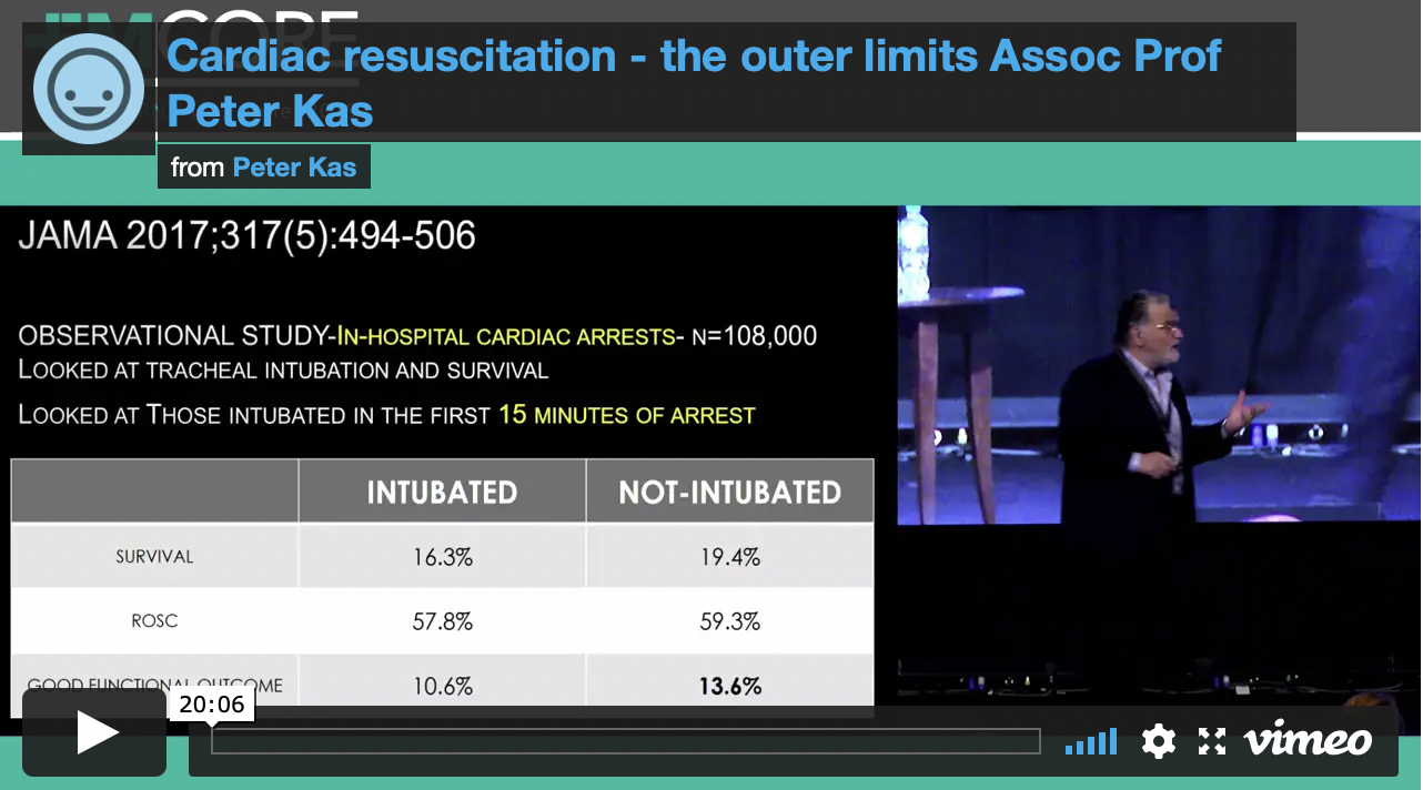
The studies in out of hospital cardiac arrest showed that intubation conferred no benefit over BVM or supraglottic airway. In in-hospital cardiac arrests, those patients intubated within the first 15 minutes of cardiac arrest, had a worst outcome.
ADRENALINE
The use of 1 mg of adrenaline which initially came from canine models, was introduced at this time and has been a major part of all resuscitations since then.
The clinical significance of adrenaline is uncertain. It’s been shown to give no improvement and even to decrease survival to hospital discharge and decrease the rate of favourable neurological outcomes. Steil in the multi-centre OPALS study demonstrated no improvement in neurologically intact survivors to hospital discharge when using advanced life-support. We know that high-dose adrenaline doesn’t improve outcomes.
Jacobs, further randomised 601 cardiac arrest patients and found a significantly improved likelihood of achieving return of spontaneous circulation(ROSC) in the adrenaline group. There was a trend to increased survival to discharge in this group, unfortunately the study was underpowered.

Everyone should get 1mg of Adrenaline right?
Giving 1mg of adrenaline to a heart that has just resumed beating, can have dire consequences. The effect of markedly increased afterload against which a weak heart must beat, can result in a rapid loss of circulation. The adrenaline dose must be titrated to achieve adequate coronary and cerebral perfusion.
Our aim should be to achieve a coronary perfusion pressure of above 15 – 20 mmHg. This equates to diastolic blood pressure of 25 – 35 mmHg, which has been shown to provide adequate cerebral perfusion pressure.
Monitoring with an arterial line is the only way to do this accurately. As I have discussed at several EMCORE conferences, the aim is to locate the femoral artery with ultrasound during the first rhythm check pause and to then pass the catheter during the second rhythm check. This requires practice and coordination of the team.
If you don’t have an arterial line, don’t trust the pulse check, or the electrical wave form. Use the cardiac ultrasound to verify whether the heart is beating or not.
In the FEEL study it was found the 38% of patients with asystole on the ECG, had coordinated cardiac motion on echo. It was further found that 58% of those in PEA had coordinated cardiac motion seen when the heart was visualised. Hypothesize what might happen with the massive alpha effects of giving 1 mg of adrenaline to these hearts. Keep the dose small if the heart is beating.
Cardiac Bootcamp Members go to the Cardiac Pearls Section for More PLUS watch the Video
Picture
JOIN THE CARDIAC BOOTCAMP COURSE
0 Comments

How would you treat this wct?

25/8/2021

1 Comment

 
Wide complex tachycardias can be a little scary. Bootcamp Members can go to the VT section and review wide complex tachycardias. Also a New section has just been added on monomorphic and polymorphic ventricular tachycardias.
This week we look at two simple cases. The key is is to pick the right treatment. The wrong treatment may simply not work, but worst than that, it may kill your patient.
CASE 1
A 32 yo male is brought into the emergency department from the methadone clinic across the road. It appears that he has been having short bursts of seizure activity. He has no other past history apart from IVDU and he is on the methadone program.
​An ECG is performed as shown below. See what you think.
Picture
He soon begins to have 'seizure' activity. You attach him to a monitor and the following rhythm strip is obtained.
Picture
What is the diagnosis?
(a) Monomorphic Ventricular Tachycardia
(b) Polymorphic Ventricular Tachycardia
(c) Torsades de Pointes
ANSWER
This is Torsades de Pointes
The definition of Torsades de Poines is a polymorphic ventricular tachycardia in a patient with prolonged QT interval. This patient had a very prolonged QT secondary to hi Methadone.
​The patient receives one shock and reverts to sinus rhythm. You wish to start medication to prevent the patient from going back into this polymorphic ventricular tachycardia.
Which one of these would you use?
(a) Procainamide

(b) Amiodarone
(c) Magnesium
​(d) All of the Above
ANSWER
The answer is (c) Magnesium.
DO NOT use Amiodarone or Procainamide as these may prolong the QT interval even further.
CASE 2
An 85 yo male presents with palpitations and pre-syncope. He has a past history of hypertension and had an AMI 5 years ago.
His cardiac examination is normal and he is neurologically intact.
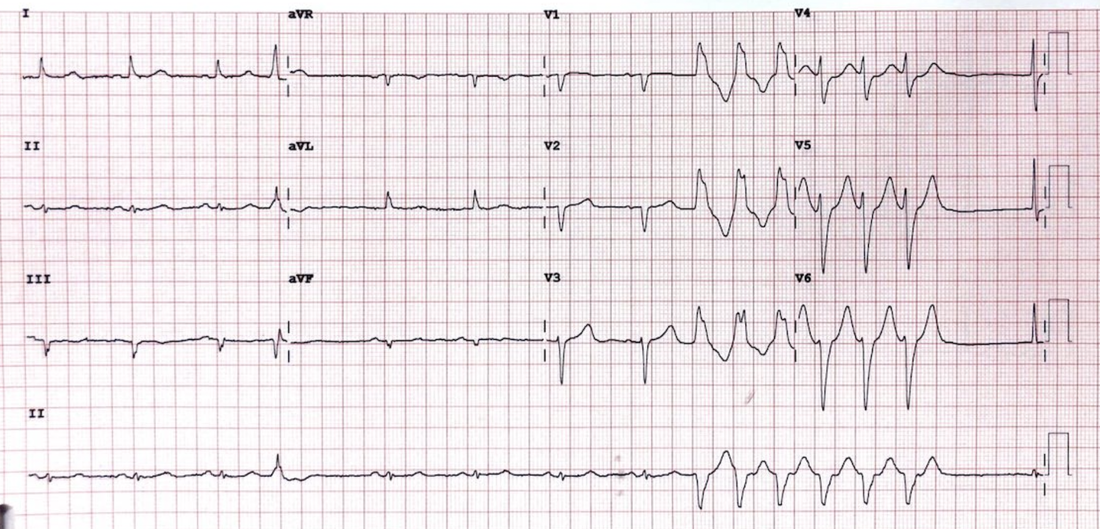
​You perform an ECG, shown below:
Picture
What is the diagnosis?
(a) Monomorphic Ventricular Tachycardia
(b) Polymorphic Ventricular Tachycardia
(c) Torsades de Pointes
ANSWER
Monomorphic Ventricular tachycardia
This is non-sustained; defined as < 3 complexes or < 30seconds.
You decide to treat the patient to prevent a recurrence. What would you use?
(a) Potassium
(b) Amiodarone
(c) Magnesium
​(d) All of the Above
ANSWER
Amiodarone can be used here. Procainamide is an alternative, however in patients with poor left ventricular ejection fraction, it can worsen heart failure.
Cardiac Bootcamp Members go to Monomorphic/Polymorphic Ventricular Tachycardia
1 Comment

A 19 yo with a puzzling post reversion ecg.

17/8/2021

0 Comments

 
A 19 yo male presents to the emergency department with palpitations. He has a narrow complex tachycardia on the monitor at a rate of 155 beats per minute. He has some left sided chest discomfort which this is pinpoint, sharp and totally reproducible. He states that he has had this for the last 2 days following working out at the gym. 
An ECG is done and you diagnose SVT. You discuss what SVT means with the patient and progress to vagal manoeuvres, which don't revert the patient. You then discuss the use of Adenosine with the patient. The patient reverts with a single dose of Adenosine.
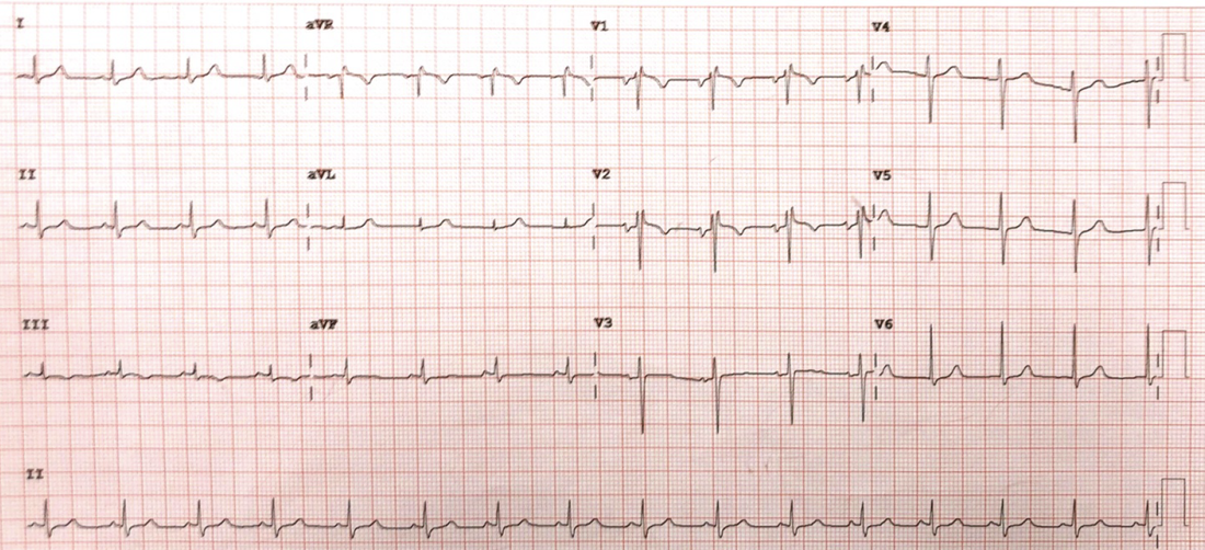
​The post reversion ECG is shown below. What does it show?
Picture
What is the Diagnosis?
(a) Anterior ST elevation Infarction

(b) Pulmonary Embolism
(c) Brugada Syndrome
(d) Lead misplacement
(e) Arrhythmogenic Right Ventricular Dysplasia
ANSWER
(d) Lead misplacement
​The ECG shows a RBBB pattern in V1 with inverted T waves in V1-2.

​The p waves are the key here. The p wave in V1 is inverted and biphasic in V2. This indicates that these two leads are placed too high.
CARDIAC BOOTCAMP COURSE MEMBERS CLICK HERE TO READ MORE
0 Comments

A 65 yo male IN cardiogenic shock: pressors?

10/8/2021

0 Comments

 
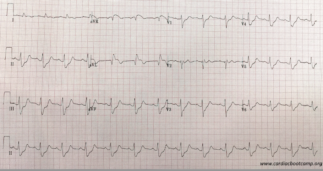
A 65 yo male is brought in by ambulance. He has had chest pain for the previous 3 hours. He is diaphoretic and looks unwell. His BP is 71/50 and he is speaking in single words, with saturations of 90% on a non-rebreather. His chest examination has widespread crepitations.
He has no relevant past medical history.
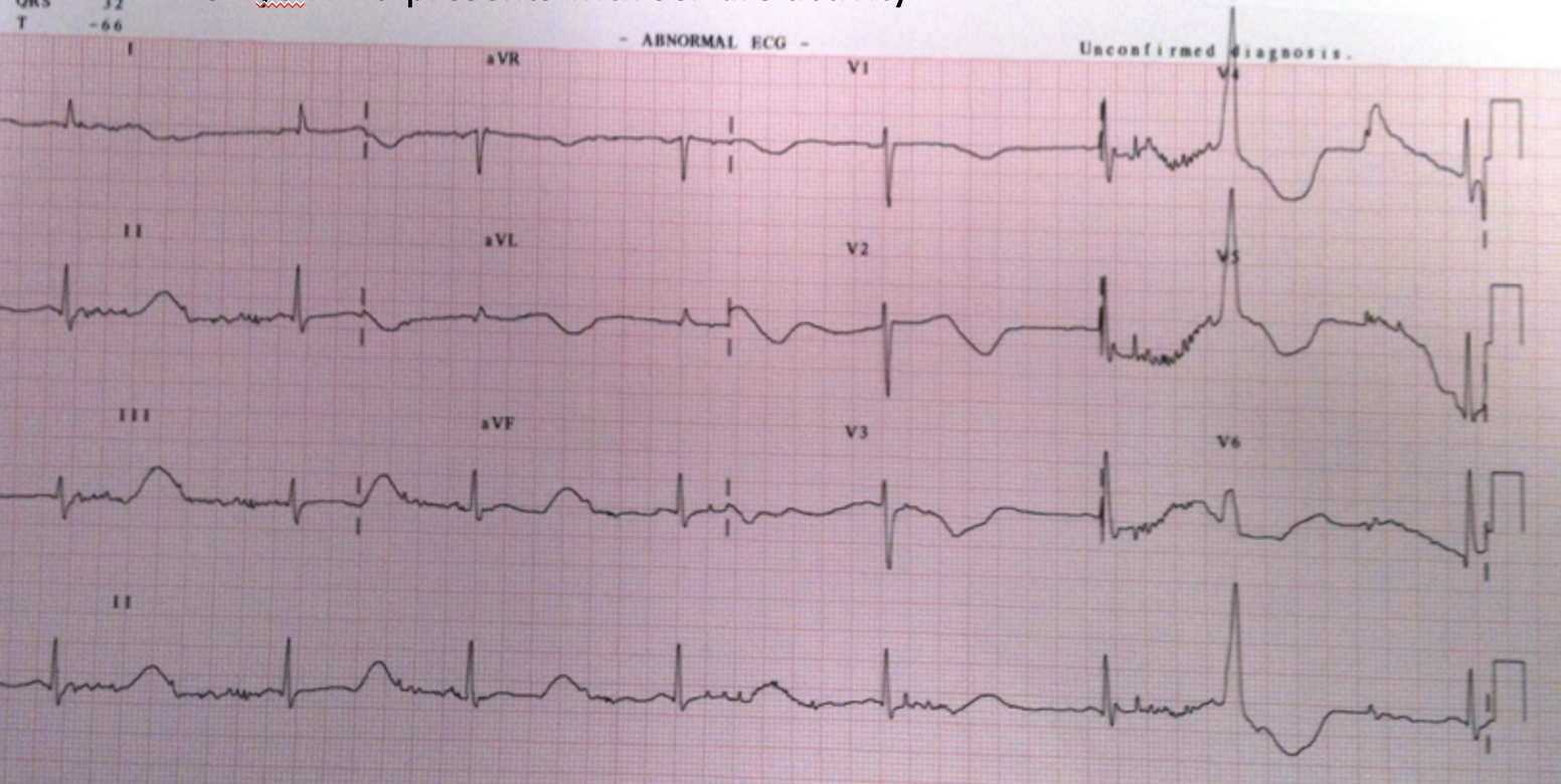
Hi ECG is shown below:

Picture
You diagnose a probable LAD occlusion and that the patient is in cardiogenic shock. Your hospital doesn't have a Cath lab and so you decide to thrombolyse. However, you also need to sort out the cardiac failure and the blood pressure.
GTN is not an option to dilate due to the cariogenic shock. You decide on CPAP for the breathing but need to support the blood pressure. What do you use?
  1. Adrenaline
  2. Noradrenaline
  3. Dopamine
  4. Isoprenaline
  5. Dobutamine
  6. Milrinone
  7. Vassopressin

​Some Important Comments in the LiteratureIs one Pressor better than the other? Perhaps another question to ask is if one pressor, although efficacious in the short term lead to worst outcome? By this I mean, something that may work well right now, but lead to worst outcomes a week or two down the track when the patient is out of the emergency department.
The Cochrane Review first published findings in 2016 of Vasopressors in hypotensive shock and said"
“We found no evidence of substantial differences in total mortality between several vasopressors. Dopamine increases the risk of arrhythmia compared with norepinephrine and might increase mortality. Otherwise, evidence of any other differences between any of the six vasopressors examined is insufficient.”
Cochrane Database of Systematic reviewsTarvasmaki et al, in an observational study in Critical Care (2016) 20:208 found that patients on Adrenaline had a worst survival and an increased 90 day mortality compared to other vasopressors. However the study didn't prove causality and a prospective trial is needed.
In a comparison of Dopamine and Noradrenaline in the treatment of shock, De Baker et al (NEJM 2010;362(9):779) found that although not significant, there was a trend towards a higher death rate when Dopamine was used. There were certainly a significant number of adverse events such as arrhythmias, when Dopamine was used. In a sub-group analysis in this same study they found that in cariogenic shock, Noradrenaline was superior to dopamine in terms of reduction in mortality.

This was the case of the 65 yo male in CARDIOGENIC SHOCK.

The optimal treatment for this patient would have been to start NorAdrenaline and then add Dobutamine, especially as there was no previous history of heart disease. Milrinone may be a substitute in patients with known poor ejection fraction ie., known significant heart failure.
The key is to start with Noradrenaline.

A quick review of the Medications available

α1 = vascular(arterial) smooth muscle
β1 = Heart: Increases rate and force of contraction
β2 = Bronchial Smooth muscle dilatation

INOPRESSORS: Adrenaline and NorAdrenaline

Adrenaline (αβββ)
Non-selective adrenergic agonist: acts on α and β receptors. At low doses, acts on β2 receptors causing vasodilation. At higher doses its effect on α1 and β1, resulting in positive inotropy and vasoconstriction( of peripheral vasculature and pulmonary arterial and venous circulation).
It is both a chronotrope and an inotrope
It's use is mostly in anaphylaxis and cardiac arrest as other drug combinations have been found to be better for both septic and cariogenic shock.

NorAdrenaline(αααβ)
It acts on vascular α1 adrenergic receptors, causing vasoconstriction and thus increasing systolic and diastolic blood pressures. It has chronotropic and inotropic effects by acting on on cardiac α1 receptors.
It can cause tachycardia and increase myocardial oxygen demand. It may also have a direct toxic effect on cardiac cells. It is the first line management of septic shock and may be used with other medications.

INODILATORS: Dobutamine and Milrinone

Dobutamine (αββ)
Dobutamine, a synthetic catecholamine, acts on β1 receptors increasing cardiac contractility. It also has α1 and β1 effects on peripheral vasculature resulting in vasodilatation at lower doses, thus increasing cardiac output. At doses above 5mcg/kg/min it can result in vasoconstriction.
It is usually used with an inopressor in cariogenic shock.
A meta-analysis, showed Dobutamine to be associated with higher in-hospital mortality and readmission rates for heart failure exacerbation when compared to nesiritide therapy. Higher doses of dobutamine is not preferred in patients with recent myocardial ischemia, as it can increase myocardial oxygen demand and induce tachycardia.

Milrinone
It is a Phosphodiesterase Type 3 inhibitor(PDE3) used inotropic agent in patients with cardiogenic shock. It inhibits PDE3, which results in more calcium ions entering the myocardial cell, increasing cardiac contractility. It also acts on peripheral and pulmonary vasculature leading to vasodilatation as well as inotropic effect. It does not act on the beta adrenergic pathway.
Another feature of the mechanism of action of milrinone is that the same intracellular processes is activated in smooth muscle cells of the peripheral and pulmonary vasculature, leading to a net vasodilatory effect in addition to its positive inotropic effect.
In a sub-group analysis of the OPTIME-CHF trial, it was found to increase mortality in patients with heart failure of ischaemic origin.

Conclusion:
Think about the medications you are using in cardiogenic shock.
Start with Noradrenaline and then add to that. In most cases it will be Dobutamine.
0 Comments

A 60 yo with chest pain.... deteriorates

2/8/2021

0 Comments

 
This blog is based on a case study in the literature.
A 60 yo male with a recent previous history of angina (with a cardiac stent), has intermittent left sided chest pain. His past medical history is Hypertension and Diabetes. He is haemodynamically stable with a heart rate of 93bpm and Blood Pressure of 122/80 and a respiratory rate of 18.
His ECG shows Q waves in leads III and aVF and his troponin is normal.

4 hours later he crashes,  becomes diaphoretic and tachypnoeic, with a heart rate of 136 bpm and a BP of 70mmHg.
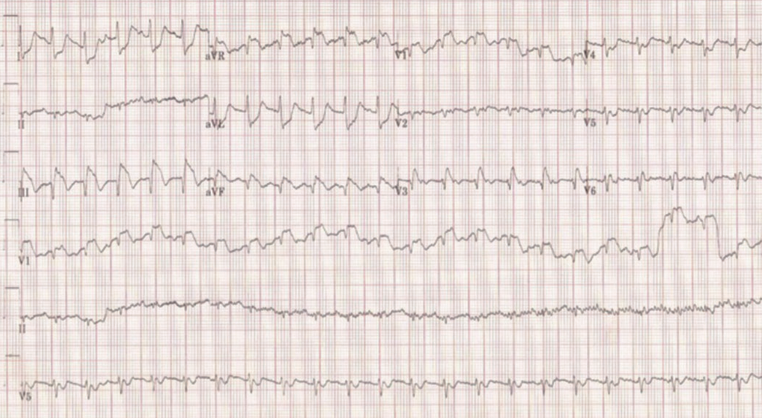
An ECG is done and shown below. Based on this, the patient is taken to the cath lab.
​What does it show? What is the diagnosis?
Picture
Source: Vascular 2019
ANSWER
  • Sinus tachycardia
  • ST elevation in III, aVF and aVR and V1 with reciprocal changes in precordial leads and lateral leads. The ST elevation and reciprocal changes are consistent with a STEMI
​Can you see anything else?
.... There is an S1Q3T3
There was no coronary artery obstruction was found.
​A CTPA was done that revealed a large saddle pulmonary embolism.
​
​Let's look at PE mimicking an acute infarction.

Clinical Presentation of PE

Match the following percentages with the following symptoms:
79%,   57%,   47%,   26%
  1. Pleuritic chest pain
  2. Tachycardia
  3. Tachypnoea
  4. New dyspnoea at rest or exertion
ANSWER
  1. Pleuritic chest pain. 47%
  2. Tachycardia. 26%​
  3. Tachypnoea. 57%
  4. New dyspnoea at rest or exertion. 79%

What are some of the ECG Findings in PE?

Findings on ECG, for the most, are non-specific for diagnosing pulmonary embolism.
Some of the more typical findings include: 
  • Sinus Tachycardia
  • S1Q3T3
  • S1Q3
  • Right Axis
  • Bundle Branch Block (complete or incomplete)
  • T wave inversions in the right precordial leads (right heart strain).
Is st elevation a typical ecg finding in pulmonary embolism?
Not normally. However if it is present, it is usually in anteroseptal leads rather than inferior leads and is least likely to be present in the lateral leads.

Other ECG abnormalities

In a study by Sreeram et al, it was found that > 3 of the following increased probability of PE:
  • RBBB (complete or incomplete) + STE and positive T wave in V1
  • Right axis deviation
  • S waves >1.5mm in I and aVL
  • Shift of transition zone in precordial leads to V5
  • Q waves in III and aVF (but not II)
  • T wave inversion in III, aVF, V1-4
  • Low voltage QRS in limb leads of <5mm
why is there a troponin leak in pulmonary embolism?
Troponin leaks can occur from myocardial stretching secondary to right ventricular afterload increases.

It's hard to remember everything

You can't remember everything, so look these up next time a patient presents. The ECG must be looked at with the patient's presenting complaint and past history.
I remember  to look for 5 things:
  1. Unexplained Sinus Tachycardia
  2. S1Q3T3 or S1Q3
  3. RBBB (complete or incomplete) + right axis deviation
  4. T wave inversion in III and V1-4
  5. S wave in I (+ aVL)
Member, find out more by going to the pulmonary embolism section
Topics on Pulmonary embolism in the members section include:
  • The ECGs of Pulmonary Embolism
  • ECG findings that predict your patient is about to crash
  • What are the symptoms and clinical signs of pulmonary embolism?
  • Scoring systems
  • Investigations such as the ABG and D-dimer
  • PE in Pregnancy
  • What about sub-segmental PE's?
  • Who needs thrombolysis?
  • The most important studies in management of PE

REFERENCES
  1. Villablanca P A et al. Case report and systematic review of pulmonary embolism mimicking ST-elevation myocardial infarction. Vascular 2019, Vol 27(I) 90-97.
  2. Falterman TJ, et al. Pulmonary embolism with ST segment elevation in leads V1 to V4: case report and review of the literature regarding electrocardiographic changes in acute pulmonary embolism. J Emerg Med 2001; 21: 255–261. 
  3. Sreeram N, et al. Value of the 12-lead electrocardiogram at hospital admission in the diagnosis of pulmonary embolism. Am J Cardiol 1994; 73: 298–303. 


0 Comments

    Author

    Dr Peter Kas
    ​Emergency Physician

    Back to resus.com.au
    Picture
    Picture
    Sign up for
    ECG of the week
     Its free

    Archives

    May 2023
    March 2023
    February 2023
    January 2023
    December 2022
    November 2022
    October 2022
    September 2022
    August 2022
    July 2022
    June 2022
    March 2022
    December 2021
    November 2021
    October 2021
    September 2021
    August 2021
    July 2021
    June 2021
    May 2021
    April 2021
    March 2021
    February 2021
    January 2021
    December 2020
    November 2020
    September 2020
    August 2020
    July 2020

    Categories

    All
    Ablation
    Atrial Fibrillation
    Atrial Flutter
    Atrial Tachycardia
    Axis
    Bradycardia
    Cardiac Arrest
    Chest Pain
    ECG Mimics
    Ischaemia
    Lead Reversal
    Migraine
    Myopericarditis
    Pacing
    Paediatric Arrhythmias
    Paediatric ECG
    Paediatric Syncope
    Pericarditis
    QT Interval
    STEMI
    STEMI Mimics
    SVT
    T Waves
    Ventricular Tachycardia
    Wellens
    Wide Complex Tachycardia

Our Other Websites to Visit

www.emcore.com.au
www.resus.com.au

Contact Us
© COPYRIGHT 2015. ALL RIGHTS RESERVED.
  • HOME
  • Course Login
    • Log In
    • Log Out
  • SYLLABUS
  • STORE
    • Online Self Study Course
    • Cardiac Video Course
  • ECG OF THE WEEK