CARDIAC BOOTCAMP
  • HOME
  • Course Login
    • Log In
    • Log Out
  • SYLLABUS
  • STORE
    • Online Self Study Course
    • Cardiac Video Course
  • ECG OF THE WEEK

ECG OF THE WEEK

BECOME A COURSE MEMBER
Each Week we put up one ECG case for you....because it's easier to learn from cases.
The ECG of the week is FREE to everyone. To become an expert at ECG's join the Cardiac Bootcamp Course. It's a great way to Learn!
​Or come to one of our LIVE Cardiac Bootcamps

An 81 yo male with chest pain

30/7/2021

1 Comment

 
It's mid morning in the emergency department.
An 81 yo male presents to triage with chest pain. The pain started last evening ie., 14 hours previously and is still present.
The pain is described as a pressure pain with some radiation into the jaw area. There is no diaphoresis, no dyspnoea and no distress.
There is minimal past medical history.
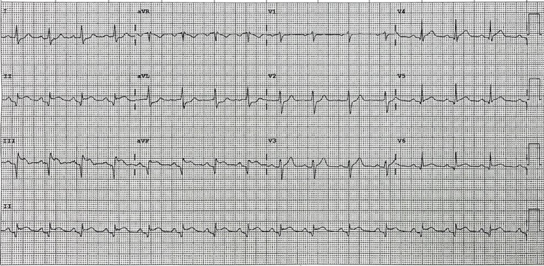
​An ECG is done. Take a moment to look at it. 
QUESTION: 
What does it show?
Picture
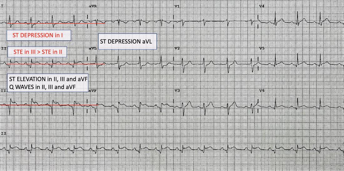
ANSWER: There is an INFERIOR infarct. In most cases this is due to a RIGHT CORONARY ARTERY occlusion but in a small number of cases a left circumflex artery occlusion can also  cause this.  
​A right coronary occlusion is most likely as when the following are present:
  • STE in III > STE in II and
  • there is some ST depression in I (see below).
​If this were a circumflex lesion, these  changes would not have occurred.
​
The patient gets ASPIRIN and you prepare to give GTN.

QUESTION: Should you give GTN? What ECG should you perform before this?
Picture
ANSWER: We know that up to half of all patients with an inferior infarction will develop RIGHT VENTRICULAR infarction. Right Ventricular infarction is associated with a greater than two-fold increase in mortality as well as an increased risk of arrhythmias and  high grade AV-nodal blocks.
Patients with a right ventricular infarction, are preload dependant ie., if we drop the preload, by giving a dilator such as GTN, they drop their blood pressure significantly. If you do give them GTN without looking at the Right Ventricular leads, they need IV fluid pushed, to improve the preload and they may need inotropes.

​In a 
RIGHT VENTRICULAR INFARCTION  the following are present on the ECG
  • STE in III > STE in II and
  • ST elevation in aVF > ST depression in V2.
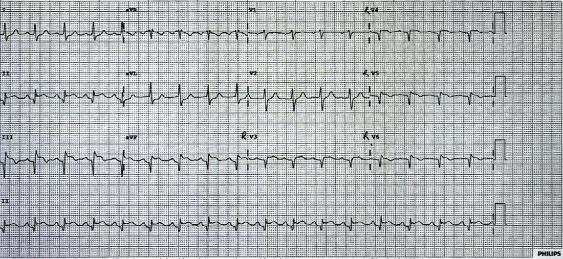
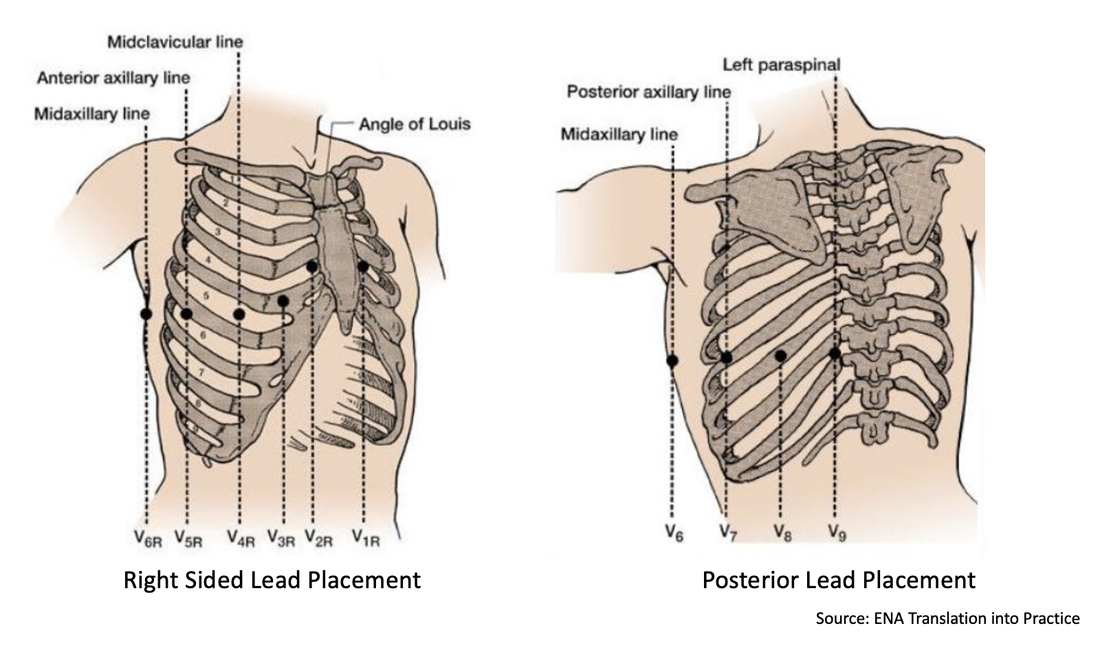
Right Ventricular leads were performed on this patient. See below. V4 is now right sided V4. RV4 is obtained by looking at the right side of the heart and placing V4 in the right mid-clavicular line in the 5th intercostal space.
ST elevation in RV4 has a 100% sensitivity and an 87% specificity and 92% predictive accuracy in diagnosing right ventricular myocardial Infarction.
​

BEWARE: In up to 50% of patients with a Right Ventricular Infarction the ST Elevation may disappear after about 12 hours. If there is no right ventricular ST elevation, but the patient decompensates following nitrates, suspect it.
Picture
Right Ventricular Leads

​What occurs in Right Ventricular Infarction?

Why are patients preload dependant and why do they drop their blood pressure?
With a right ventricular infarction, the right ventricle cannot pump properly and the right ventricular volume and end-diastolic pressure of the right ventricle are high. Due to the pump failure, the left ventricle is not receiving a sufficient amount of blood, but now the right ventricle with it's increased volume also displaces the septum towards the left ventricle, decreasing its volume even further..
Anything that decreases preload ie., diuretics nitrates, beta blockers, morphine, can significantly decrease blood pressure. These patient may then require IV fluids and inotropic support.

​POSTERIOR Myocardial Infarction

Up to 20% of inferior infarctions will present as a posterior infarction. This represents a significantly larger area of infarction. The diagnosis is made by performing a posterior lead ECG ie leads V7-9. Some clues of a posterior infarction on the normal ECG include:
  • Horizontal ST depression in V1-3
  • Dominant R wave in V2
  • Upright T waves.
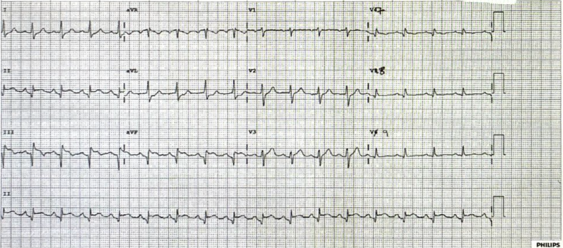
The initial ECG showed some mild ST depression in V2, but little else. Posterior leads were performed, as shown in the ECG below.
Picture
Posterior Leads
There are no posterior changes in leads V7-9

​OUTCOME

The patient was transferred out for catheterisation and diagnosis. A 100% right coronary artery occlusion was stented.

QUESTION: Could we have given thrombolytics?
Time from symptom onset, is a marker of downstream myocardial damage. Usually late presentations(>12 hours) are referred for percutaneous coronary intervention, although not always. Three studies (references given below) a worth reading here:
  1. The LATE Study: No mortality benefits using thrombolytics in those patients presenting > 12 hours
  2. BRAVE-2 Trial: Prognostic benefit in recanalisation in late presenters up to 48 hours.
  3. OAT Trial showed no improvement in mortality with PCI in very late presenters

My approach is that given that the patient was stable and had presented more than 14 hours following symptom onset, transfer for PCI was appropriate. If there was no cath lab and there would be a significant delay in treatment, I would discuss thrombolysis with the Cardiologist. Rescue plasty could always occur later.

All Cardiac Bootcamp Self Study Course Members should review the topic 'Subtle Ischaemic Changes', in the syllabus, especially changes in aVL.

How to set up Right Sided and Posterior ECG Leads

Picture

REFERENCES
  1. Hamon M, et al. Prognostic impact of right ventricular involvement in patients with acute myocardial infarction: meta-analysis. Crit Care Med. 2008 Jul;36(7):2023–33. 
  2. Somers MP, Brady WJ, Bateman DC, Mattu A, Perron AD. Additional electrocardiographic leads in the ED chest pain patient: right ventricular and posterior leads. Am J Emerg Med. 2003 Nov;21(7):563–73.
  3. Shiraki H, Yokozuka H, Negishi K, et al. Acute impact of right ventricular infarction on early hemodynamic course after inferior myocardial infarction. Circ J. 2010 Jan;74(1):148–55.
  4. . Late Assessment of Thrombolytic Efficacy (LATE) study with alteplase 6–24 hours after onset of acute myocardial infarction. Lancet. 1993; 342:759–766.
  5. Ndrepepa G, et al. Mechanical reperfusion and long-term mortality in patients with acute myocardial infarction presenting 12 to 48 hours from onset of symptoms. JAMA. 2009; 301:487–488.
  6. Hochman JS, et al. Occluded Artery Trial Investigators. Coronary intervention for persistent occlusion after myocardial infarction. N Engl J Med. 2006; 355:2395–2407.
1 Comment

The Patient with syncope: ECG Quiz

20/7/2021

0 Comments

 
This week I am putting on a 30 minute webinar on The Approach to the patient with Syncope.
Date: Thursday July 22nd at 3.00pm (Melbourne Time)
Here is the Zoom link: 
​https://us02web.zoom.us/j/87324913445?pwd=VkwwcHhjdHZJd09JT2N3c214RDVKdz09

Before the Webinar Try your hand at these few ECGs.

The ECG has a < 5% yield but is still the highest yield of all investigations we can do in the syncope patient. It's importance cannot be understated,  as cardiac causes of syncope have the highest mortality and morbidity of all causes.

Below are 7 ECGs of patients that present with syncope

What is the diagnosis in each case?

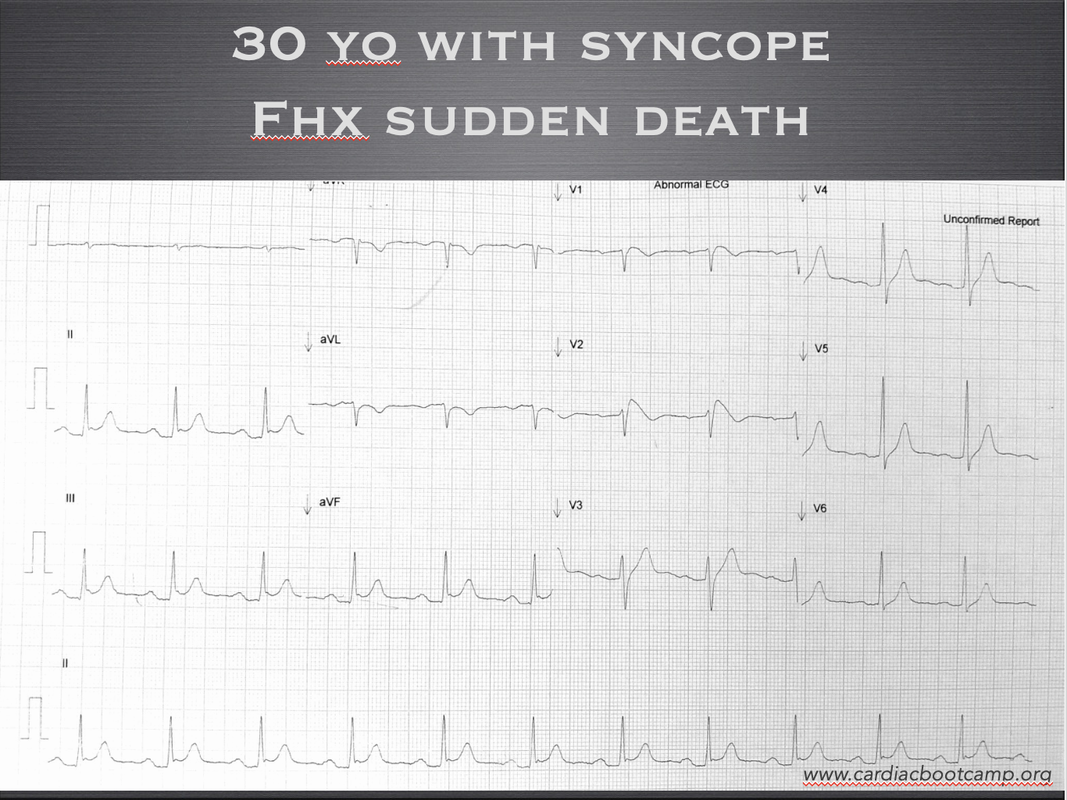
ECG 1: 

A 30 yo male presents with his third episode of syncope. He was waiting for a bus and felt a brief episode of palpitations then found himself on the ground. He has no past medical history by states that his father died suddenly of unknown causes.
He has had a cardiac workup during a previous presentation, which included monitoring,  serial troponins and an echocardiogram, which were all normal.
What does the ECG show?
Picture

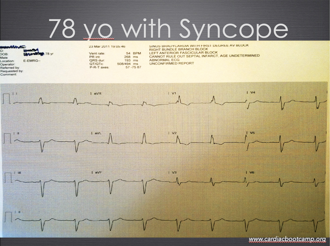
ECG 2

A 78 yo male is brought in by ambulance following a fall at home. He has no memory of the incident. He states that he was making a cup of tea, felt dizzy and then found himself on the floor of his kitchen. He couldn't get up and called for help. His wife called the ambulance.
What does his ECG show?
What probable definitive treatment will he need?
Picture

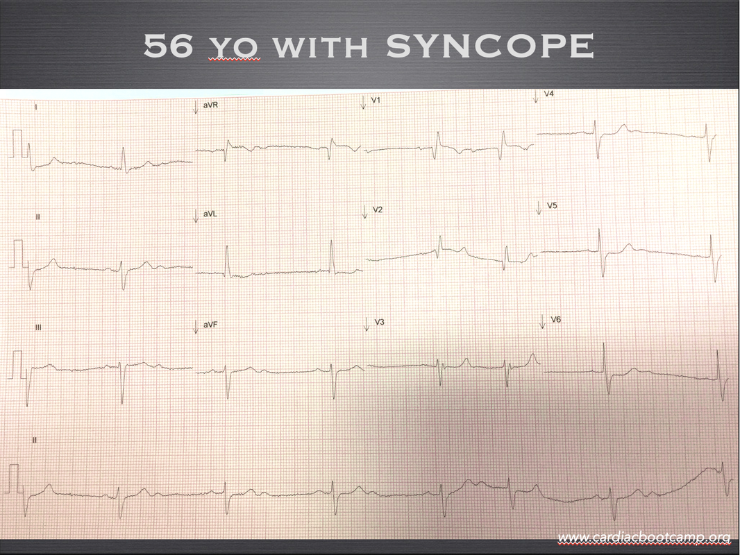
ECG 3

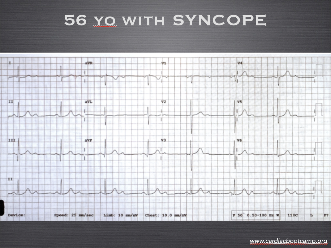
A 56 yo male presents with a syncopal episode. He had momentary dizziness, but no shortness of breath or palpitations or chest pain.
What does the ECG show and what caused his syncopal episode?
Picture

ECG 4

A 56 yo woman has presented to the emergency department following a brief syncopal episode. The patient had no chest pain, or palpitations. Following the ECG and serial normal troponins, the patient was discussed with the senior as a possible discharge.
​The senior wanted cardiology called and an admission. What was missed?
What is the diagnosis?
Picture

ECG 5

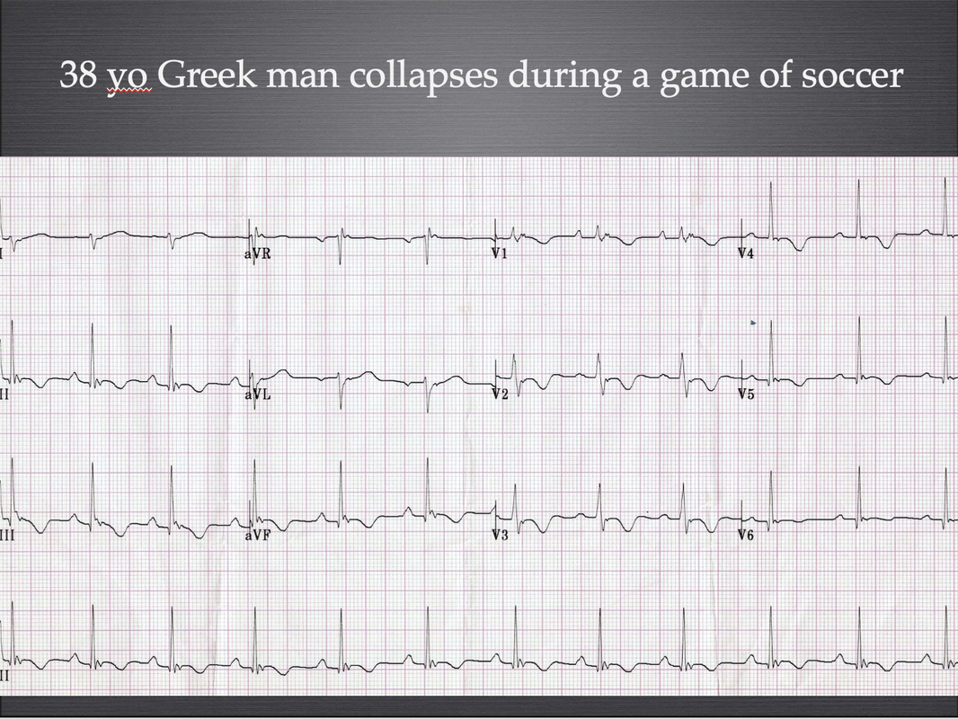
A 38 yo greek man is brought in by ambulance as he collapsed, whilst playing soccer. He has no past medical history and has had some previous episodes of palpitations in the past.
He is well and wishes to go home.
What does the ecg show?
​What waveform is shown in leads V1 and V2?
Picture

ECG 6

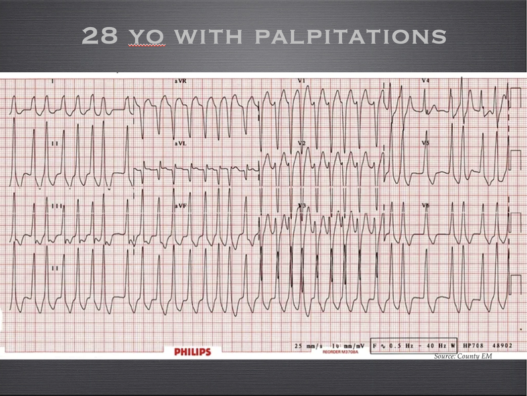
A 28 yo woman presents with palpitations and a syncopal episode. She is haemodynamically stable and looks well. There has been a history of syncope.
What is the diagnosis?
What is the treatment, or even more precisely, what medications can't we give?
Picture

ECG 7

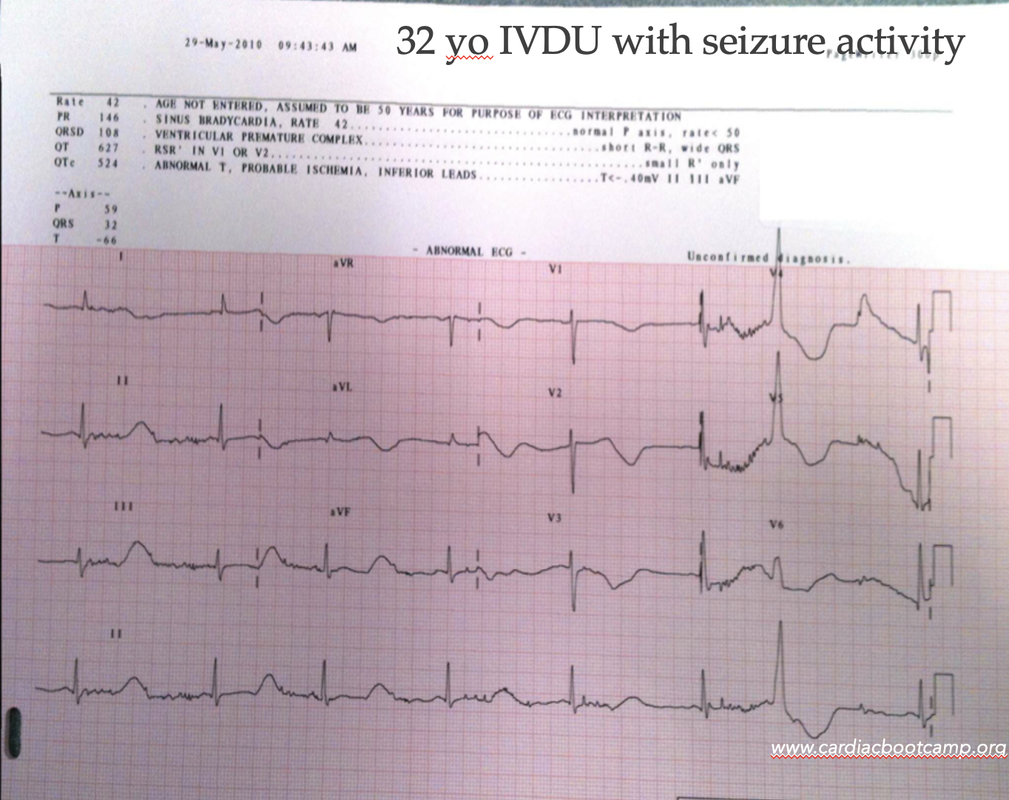
A 32 yo male is brought into the emergency department with a diagnosis of seizures. He has had several small seizures according to his partner.
He is on the methadone program.
His ecg is shown, what is the diagnosis?
​Is there a reason related to the ecg that could describe the seizures?
Picture

Try all of these. Join the zoom webinar this week. If you can't

​ make it, don't worry, I'll put up the video here next week.

0 Comments

A 28 yo patient with palpitations

13/7/2021

1 Comment

 
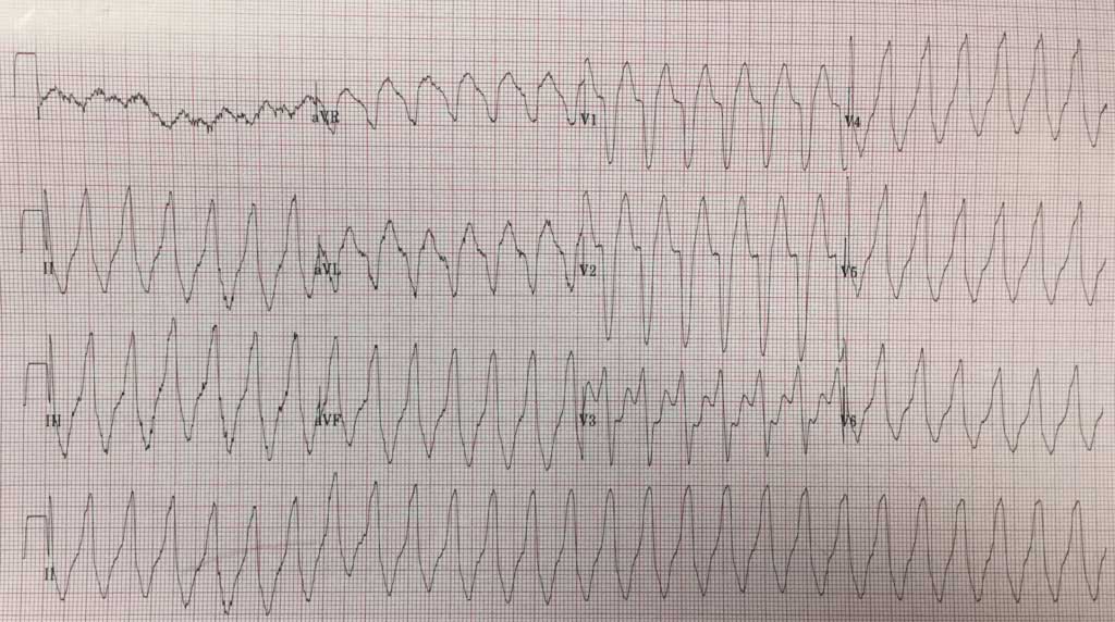
A well looking woman in her late twenties presents complaining of palpitations. She has had a past medical history of bigeminy, for which she is on metoprolol and magnesium.
She presents complaining of palpitations different to the ones she normally has with her bigeminy. She is well looking, afebrile, with a blood pressure of 142/70 and a normal respiratory rate. A Venous Blood Gas shows a potassium of 3.3mmol/L. Her ECG is as below.
What is the Diagnosis and how would you Manage this patient?
Picture
ECG INTERPRETATION
It is a wide complex tachycardia. The rate is about 1`50bpm and I think I can see 'p' waves in lead I. In a well young person, we might assume it's SVT. Could it be VT? It could be.
The patient is young so SVT with aberrancy is probably most likely. However we need to be a little bit careful with that. Ventricular  bigeminy can lead to arrhythmias.
The patient is stable and looks well, so we have some time to think.

IS IT VENTRICULAR TACHYCARDIA?
We know that 80% of wide complex tachycardias are Ventricular Tachycardia(1). There are well known rules that we can use. Let's see what happens if we use them.

BRUGADA CRITERIA(2)
The Brigade Criteria are 92% sensitive and 65% specific, so not perfect. There are 4 parts:
  1. Is there an absence of an RS complex in all precordial leads? NO
  2. R-S Interval >100ms in any precordial lead? YES- indicating VT, so we can stop here.
    1. ​Why is the RS interval important? It tells us that there is a delay in the impulse propagating, which occurs if it passes through the ventricle instead of the AV node.
VERECKEI CRITERIA(3)
The Vereckei Criteria have similar sensitivity to Brugada. Let's apply Vereckei:
  1. Is there AV Dissociation? NO
  2. Is there a dominant R wave in aVR? YES  indicating it is VT
GRIFFITH CRITERIA.
Griffith Criteria having about the same sensitivity, but rule in SVT.

It’s all too complex. In the real world cardiologists reviewing these eggs can’t agree.

WHAT IF WE ASSUMED IT WAS SVT AND GAVE ADENOSINE?
If it's ventricular tachycardia, apart from a particular kind of ventricular tachycardia, then nothing would happen, as Adenosine blocks the AV node and Ventricular tachycardia comes form the Ventricles. You wouldn't harm the patient unless it were an irregular wide rhythm (ie., AF with WPW)

We need a simple solution to working this out. We need a solution that you can apply when you’re woken at 2 o’clock in the morning.
MY 3 STEP APPROACH
  1. Is the QRS wider than 120ms(3 small squares)?
  2. Is the Rate greater than 120?
    1. The rate must be greater than 120 beats otherwise think of 3 other differentials”
      1. Hyperkalaemia
      2. Na Channel Blockade
      3. Accelerated Idioventricular Rhythm.
  3. Is this real? i.e. is it not artefacts etc.
If the answer is yes to all 3 questions, go down the VT route.  Use electricity. 200J on this patient following consent and sedation.

What would I do in this case?
Given the patient is stable and looks well, I would try a vagal manoeuvre, either the 'REVERT' technique, or the 'Reverse Vagal Manoeuvre'. If these were not successful, I would advise the patient on the side effects of Adenosine and try Adenosine. If the blood pressure dropped at any point I would prepare for cardioversion, but would give a small dose of Metaraminol, as this sometimes reverts SVT.

Always check the POTASSIUM in any arrhythmia

A final word about bigeminy. I recently wrote a blog on this topic on resus.com.au titled ‘Is Bigeminy Benign?‘ The bottom line is that we normally don’t need to do anything for these patients, as ectopics are a normal physiological response.

HOWEVER:
  1. In patients with ischaemic heart disease, these may be significant and caused by the ischaemia. Beware the multiple premature ventricular ectopics in MI.
  2. Beware premature ventricular contractions(PVC’s) and Long QTc as this may predispose to R on T phenomenon. The premature complex may fall on the T wave resulting in Torsades de Pointes.
References
  1. Zipes D P et al. Circulation 2006; 114(10) e 385-484
  2. Vereckei A et al. European Heart Journal, Volume 28, Issue 5, 1 March 2007, Pages 589–600
  3. Brugada et al Circulation Vol 83, No 5 May 1991. 1649-1659​​
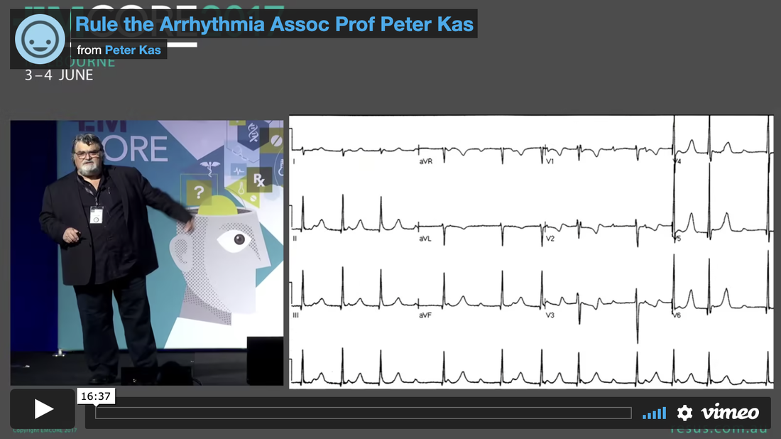
MEMBERS CLICK TO WATCH THE VIDEO RULE THE ARRHYTHMIA
Picture
1 Comment

WHAT IF WE COULD PREDICT BY EVOLVING ECG CHANGES WHO WAS GOING TO ARREST?

6/7/2021

0 Comments

 
What if we had a way to predict PEA/Asystolic Cardiac Arrest, a few minutes before it happened? Would that be helpful?.... Perhaps.
PEA and asystolic cardiac arrests have a very poor outcome. The rate of survival in out of hospital arrests is less than 2%. In hospital, survival is higher, at about 30%(1).
Having this knowledge may or may not change the outcome, but I think it gives us a significant advantage, at least in our mental preparation.

In a recent research paper by Do et al in Resuscitation(2), they identified a right ventricular strain pattern that preceded hypoxic pulseless electrical activity cardiac arrest.

STUDY DESIGN: What they did.
This was a retrospective cross sectional study at two tertiary care hospitals. They looked at in hospital cardiac arrests due to PEA/Asystole
  • Patients were at least 18 years of age
  • There was telemetry data for at least 3 hours prior to the arrest.
  • n=140
Patients were excluded if they had:
  • pulmonary hypertension
  • right ventricular enlargement on ECG,
  • ventricular paced rhythm,
  • an out of hospital arrest leading to the current admission,
  • this was not their first in hospital cardiac arrest.

Right Ventricular strain pattern
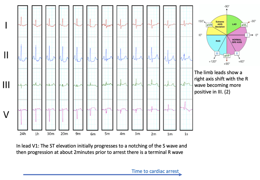
The most common terminal morphology(67% of cases) was a RBBB in V1, which progressed from an increasing RV delay as shown below:
Picture
Definite Right Ventricular strain(RVS) pattern was defined as having the above progressive morphological changes in lead V1 and either no intraventricular conduction delay or terminal intraventricular conduction delay (ie., incomplete RBBB) and two or more of the following occurring simultaneously:
  1. ST elevation V1
  2. Rightward directed ST elevation vector in limb leads (i.e. towards lead III) or
  3. Right axis deviation in limb leads
Possible RVS was defined as the RV delay pattern in V1 and only one of the supporting features.

Results
  • The median time of RVS pattern to appear on the ECG in those that had a previously normal baseline was 7.2 minutes before arrest.
  • The RVS pattern prior to PEA/Asystolic arrest was present in 47% of patients, without pre-existing right ventricular hypertrophy
  • In those patients with RVS pattern who went on to have a cardiac arrest and then ROSC, there was a 41% incidence of RV dysfunction on echo.
​
ECG Patterns identified
Picture
Conclusion
Next time we are monitoring a patient and see the gradual Right ventricular delay pattern, indicative of right ventricular failure, we should get ready as up to about half of these patients may have a PEA/Asystolic arrest in the next few minutes.

REFERENCES
  1. Holmberg Mathias J et al. Annual incidence of adult and paediatric in-hospital cardiac arrest in the United States. Circ Cardiovasc Qual Outcomes 2019; 12:e005580.
  2. Do D H. et al. Electrocardiographic right ventricular strain precedes hypoxic pulseless electrical activity cardiac arrests; Looking beyond pulmonary embolism. Resuscitation151(2020) pp 127-134.
0 Comments

    Author

    Dr Peter Kas
    ​Emergency Physician

    Picture
    Picture
    Sign up for
    ECG of the week
     Its free

    Archives

    February 2023
    January 2023
    December 2022
    November 2022
    October 2022
    September 2022
    August 2022
    July 2022
    June 2022
    March 2022
    December 2021
    November 2021
    October 2021
    September 2021
    August 2021
    July 2021
    June 2021
    May 2021
    April 2021
    March 2021
    February 2021
    January 2021
    December 2020
    November 2020
    September 2020
    August 2020
    July 2020

    Categories

    All
    Atrial Fibrillation
    Atrial Flutter
    Atrial Tachycardia
    Axis
    Bradycardia
    Cardiac Arrest
    Chest Pain
    ECG Mimics
    Ischaemia
    Lead Reversal
    Myopericarditis
    Pacing
    Paediatric Arrhythmias
    Paediatric ECG
    Paediatric Syncope
    Pericarditis
    QT Interval
    STEMI
    STEMI Mimics
    SVT
    Ventricular Tachycardia
    Wellens
    Wide Complex Tachycardia

Our Other Websites to Visit

www.emcore.com.au
www.resus.com.au

Contact Us
© COPYRIGHT 2015. ALL RIGHTS RESERVED.
  • HOME
  • Course Login
    • Log In
    • Log Out
  • SYLLABUS
  • STORE
    • Online Self Study Course
    • Cardiac Video Course
  • ECG OF THE WEEK