CARDIAC BOOTCAMP
  • HOME
  • Course Login
    • Log In
    • Log Out
  • SYLLABUS
  • STORE
    • Online Self Study Course
    • Cardiac Video Course
  • ECG OF THE WEEK

ECG OF THE WEEK

BECOME A COURSE MEMBER
Each Week we put up one ECG case for you....because it's easier to learn from cases.
The ECG of the week is FREE to everyone. To become an expert at ECG's join the Cardiac Bootcamp Course. It's a great way to Learn!
​Or come to one of our LIVE Cardiac Bootcamps

A 6week old with irritability and poor feeding

21/8/2022

1 Comment

 
A 6 week old is brought to your rural emergency department with several days history of irritability and poor feeding.
On history there was poor/no antenatal care and the child was a home birth. On examination the child is afebrile, with a tachycardia and is haemodynamically stable. There is a 4/6 holosystolic  murmur on cardiac auscultation and bilateral lung crepitations.
An ecg is done and is shown below.  Please answer the following:
  1. What does the ECG show?
  2. What is the most likely diagnosis?
  3. What investigations would you perform?
Picture
WHAT Does the ECG show?
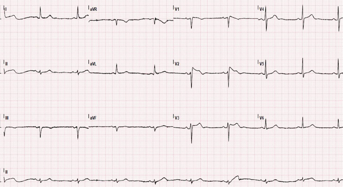
The rate is 138.
Normal P waves
The QRS's in V4-5 overlap, indicating a Left Ventricular Hypertrophy.
​This is most likely due to a Vetriculoseptal defect (VSD) 
WHAT is the most likely diagnosis
This is a VSD. VSD's cause a left to right shunt. Over time they result in pulmonary hypertension and right ventricular hypertrophy, which then reverses the shunt. This can lead to Eisenmenger's syndrome, resulting in cyanosis and heart failure.
At 4 to 6 weeks, the features of heart failure begin to appear.
what investigations would you like to order?
The diagnosis has almost been made clinically. We already have an ECG. We need a Chest-Xray, which may show cardiomegaly and a cardiac echo to follow.
Members please go to the paediatric ECG and Cardiac Conditions section
1 Comment

STEMI MIMICS

18/8/2022

0 Comments

 
STEMI mimics(1) are important to understand, because in taking the time to investigate the mimic, we can miss opportunity for early treatment of the real disease. Unlike STEMI equivalents, which have been covered previously, some of the mimics are not as serious.
​Here are some examples.

1. Normal ST Elevation

This can be in 2 forms, where in both the ST segment is concave:
  1. Male pattern: 1-3 mm STE in V1-V4
  2. Female pattern: 1mm STE in V1-V4
The formal definition of a STEMI as per AHA/ACC(2) is:
  • Men
    • < 40 yo: >2.5 mm ST-elevation in V2 or V3, 1 mm in any other lead
    • ​> 40 yo: >2.0 mm ST-elevation in V2 or V3, 1 mm in any other lead
  • Women: >1.5 mm ST-elevation in V2 or V3, 1 mm in any other lead

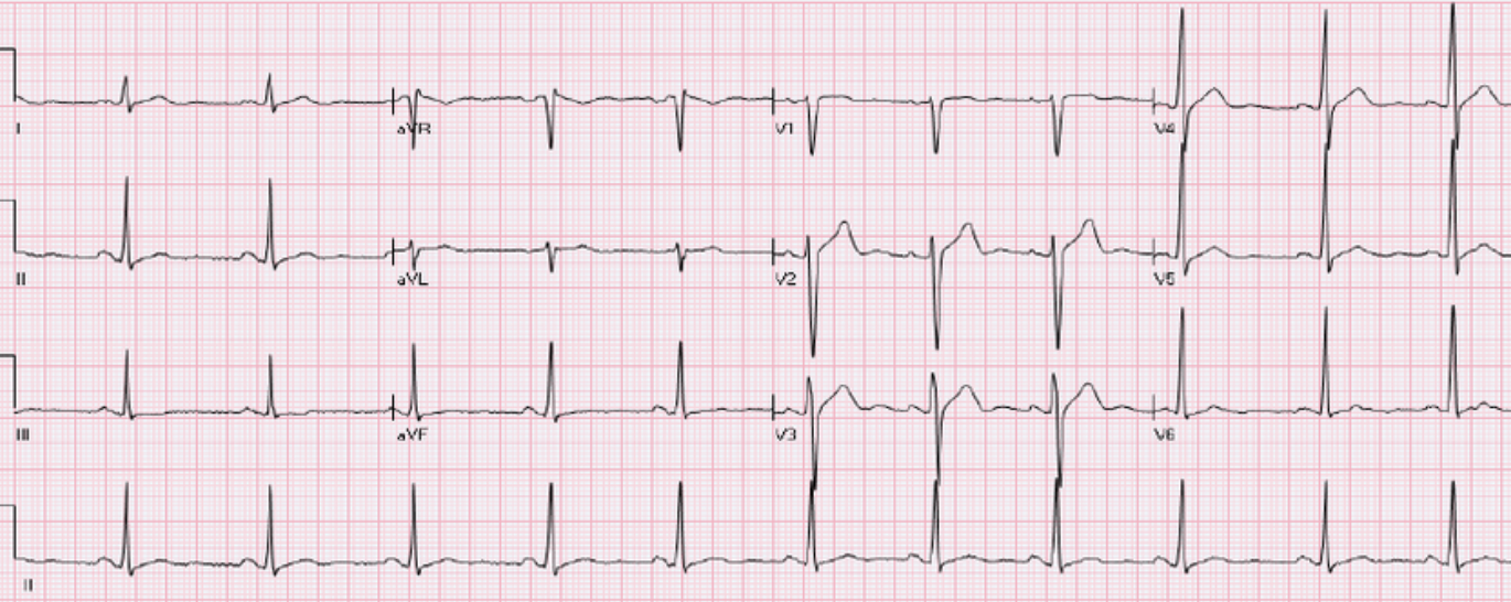
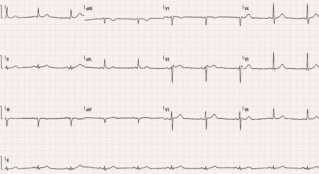
​The ECG below is from a male who presents with chest pain. It is normal.
Picture
(3)

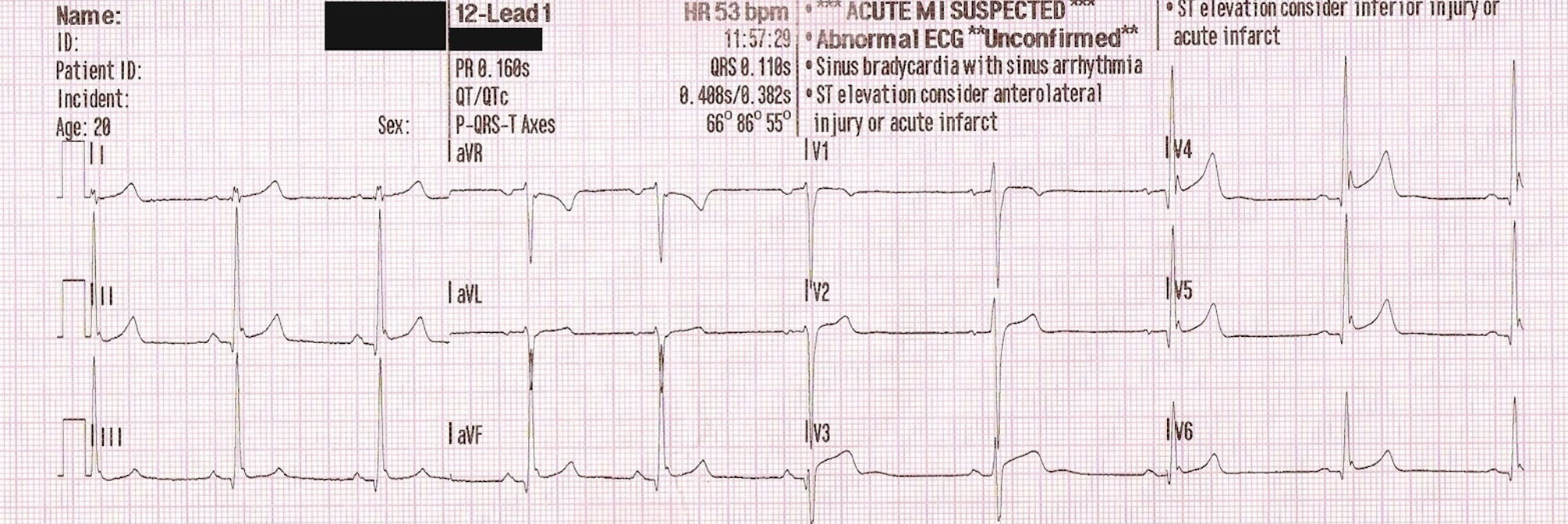
2. Benign Early Repolarisation (BER)

This condition is present in a younger population group and amongst athletes. There are three types:
  1. Type 1: Pattern occurs in the lateral leads
  2. Type 2: Pattern occurs in the inferior/inferolateral leads
  3. Type 3: Pattern occurs throughout the whole ECG.
BER Pattern:
Point notch with elevation
STE with concave up morphology V2-V6, II, III, avF usually < 2mm
No reciprocal changes
​Symmetrical Concordant T wave
​STE/T-wave height <0.25 in V6

Benign Early Repolarisation may not be that benign, being associated with serious arrhythmias in some studies. 
Picture
BER Source: Cardiac Bootcamp Online Course

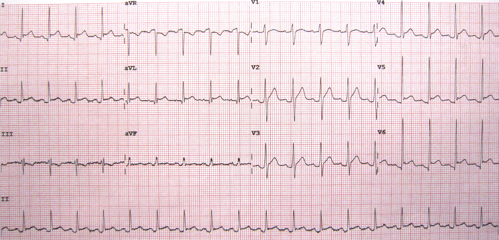
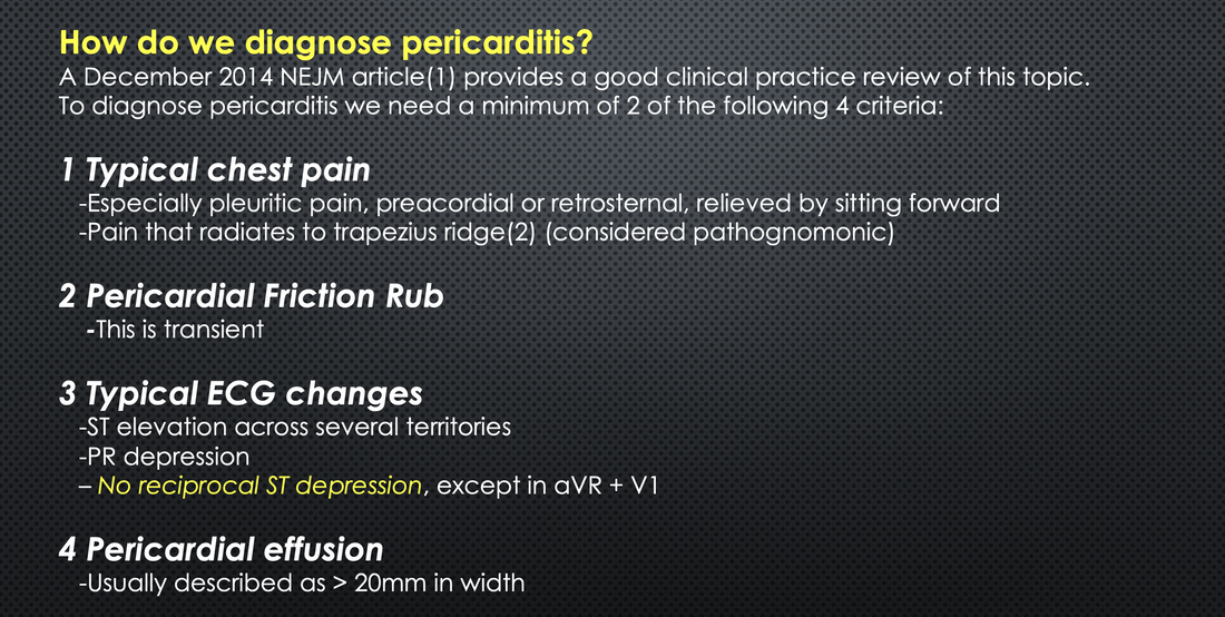
3. Pericarditis

Diffuse STE
ST/T-wave >0.25
There are 4 stages in pericarditis:
  • Stage I: ST and PR changes
    • Diffuse concave up ST segment elevation,
    • Reciprocal ST depression in  aVR. 
    • PR elevation in lead AVR + V1
  • Stage II: Normalisation of ST segments
  • Stage III: T wave inversions.
  • Stage IV: Normalisation of T waves
Picture
Pericarditis. Source: Cardiac Bootcamp Live Course
Picture

4. LBBB

ST-T and QRS discordant.
​STE is concave and <5mm

5. Hyperkalaemia

Peaked T waves
​Downsloping ST segment.

6. Brugada Syndrome

Type I: Coved Pattern
  • Leads V1-V2
  • High take-off > 2mm and concave downscoping ST segment
Picture
Type I Coved Pattern Brugada(4)
Type II: Saddle-back
  • Leads V1-V2
  • Minimum ST elevation
Picture
Type II Saddle-Back pattern Brugada(4)

7. Pulmonary Embolism

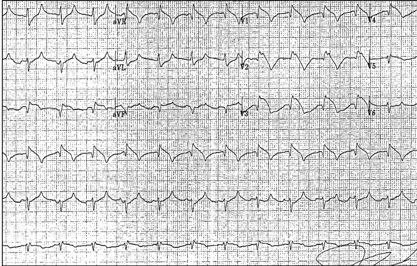
Can present with STE in Precordial leads
A more common presentation of right ventricular injury is STE in V1-V3 and/or ST depression of V4-V6.
Theories for why STE occurs in the precordial leads include:
  1. Paradoxical Embolism via atrial-septal defect or patent foramen ovale (5)
  2. Myocardial ischaemia caused by a sudden pressure load on the right ventricle, which is unable to compensate.(5)
  3. Hyperaemia from PE induces a catecholamine surge, increasing myocardial workload and ischaemia.(6)
Picture
(7)

References

  1. Wang K, et al.  ST- segment elevation in conditions other than acute myocardial infarction. N Engl J Med. 2003 Nov 27; 349(22): 2128- 35. 
  2. O'gara PT,  et al. 2013 ACCF/AHA guideline for the management of ST-elevation myocardial infarction: a report of the American College of Cardiology Foundation/American Heart Association Task Force on Practice Guidelines. Circulation. 2013;127(4):e362-425.
  3. ​Kayanı WT et al. ST Elevation: Telling Pathology from the Benign Patterns. Global Journal of Health Science. May 2012, ​4(3):51-63
  4. Mirijello A et al. Brugada electrocardiographic findings in an 80-year-old man. BMJ Case Reports. July 2013.​
  5. Cheng TO. Mechanism of ST-elevation in acute pulmonary embolism. Int J Cardiol. 2005;103:221-223
  6. Falterman TJ, et al. Pulmonary embolism with ST segment elevation in leads V1 to V4: case report and review of the literature regarding electrocardiographic changes in acute pulmonary embolism. J Emerg Med. 2001;21:255-261.
  7. ​Wilson G T et al. Pulmonary Embolism Mimicking Anteroseptal Acute Myocardial Infarction. JAOA • Vol 108 • No 7 • July 2008
0 Comments

Idiopathic Ventricular Tachycardia

10/8/2022

1 Comment

 
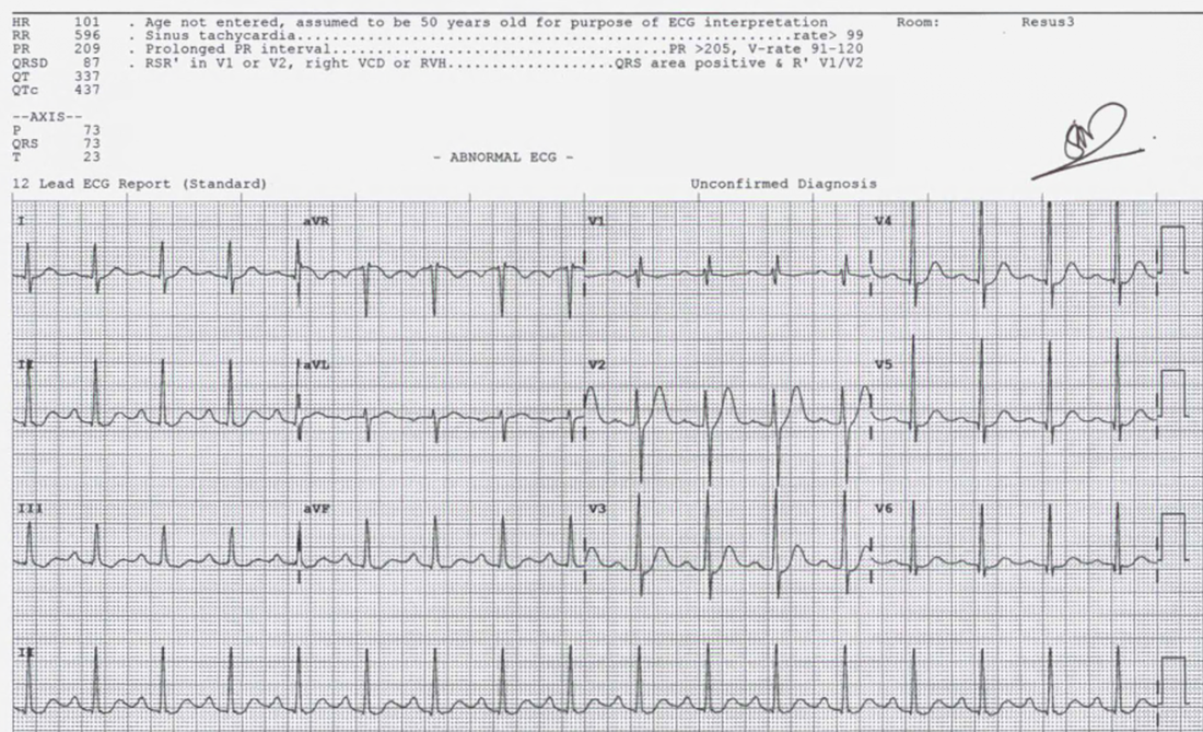
A 28 yo patient presents to the emergency department complaining of palpitations of 2 hours duration. He has no other symptoms and has no past medical history of note.
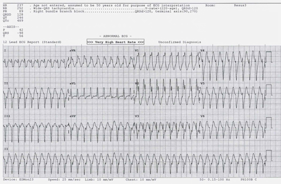
He is afebrile, with a respiratory rate of 14 a heart rate of 237bpm and a blood pressure of 119/67. 
An ECG is done and is shown below.
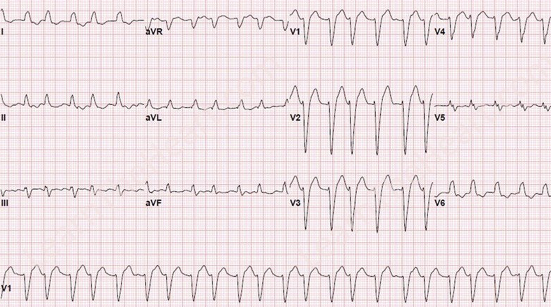
​What does the ECG show and what is your diagnosis?
​Is it Ventricular Tachycardia?
Picture
ANSWER
This is a wide 'ish' complex tachycardia at a rate of 237bpm. There is a RBBB morphology.
The axis is extreme.
If we use the 120 CRAM formula to see if it VT:
Faster than 120bpm (but not too fast)- It is faster than 120. 
Wider than 120ms- It is borderline.
Concordance: No
Initial R wave in aVR: No
AV Dissociation: No
Morphology: RBBB pattern however there is an rsR pattern not indicative of VT
R/S > 1
​
​I would consider this more likely to be Supraventricular Tachycardia (SVT) with a RBBB, even with the axis as it is.

How would you manage this haemodynamically stable patient?
(a) Adenosine
(b) Cardioversion
(c)  Sotalol IV
​(d) Amiodarone
ANSWER
This can be a difficult question to answer. For those who prefer cardioversion in every patient with a WCT, that's a very appropriate approach..
However, is there any harm in giving Adenosine? There are 3 things to consider:
  1. If this was VT, the adenosine would have no effect (unless an idiopathic VT, but we will leave that for the moment.
  2. If it is SVT with RBBB, it would hopefully revert the arrhythmia
  3. Given that the rate is very fast, could there be an accessory pathway?
The patient was given Adenosine and reverted as shown below. Was this an Idiopathic Ventricular Tachycardia. How do you differentiate it from Ventricular tachycardia or from an SVT with aberrancy?
Picture
Members go to the Idiopathic VT section for other examples and a full explanation

What is Idiopathic Ventricular Tachycardia?

Idiopathic Ventricular Tachycardia (VT), occurs in structurally normal hearts and comprises about 10% of VT cases. This is a more benign form of VT, with patients rarely suffering from sudden cardiac death.  Definitive treatment includes medications or ablation.  

Patients presenting to the emergency department with this arrhythmia, can be cardioverted and some will respond to AV nodal blockers such as Adenosine or Calcium Channel Blockers or Beta Blockers. However, even if the arrhythmia reverts with these, the patient should still be discussed with cardiology, especially if they fit the particular ECG patterns discussed below. Idiopathic VT can be mistaken for a supra ventricular tachycardia(SVT) with aberrancy and it will respond in some cases to AV nodal blockers, just like an SVT with aberrancy, so beware. However there are some distinct differences in morphology and axis, that allow us to identify it.

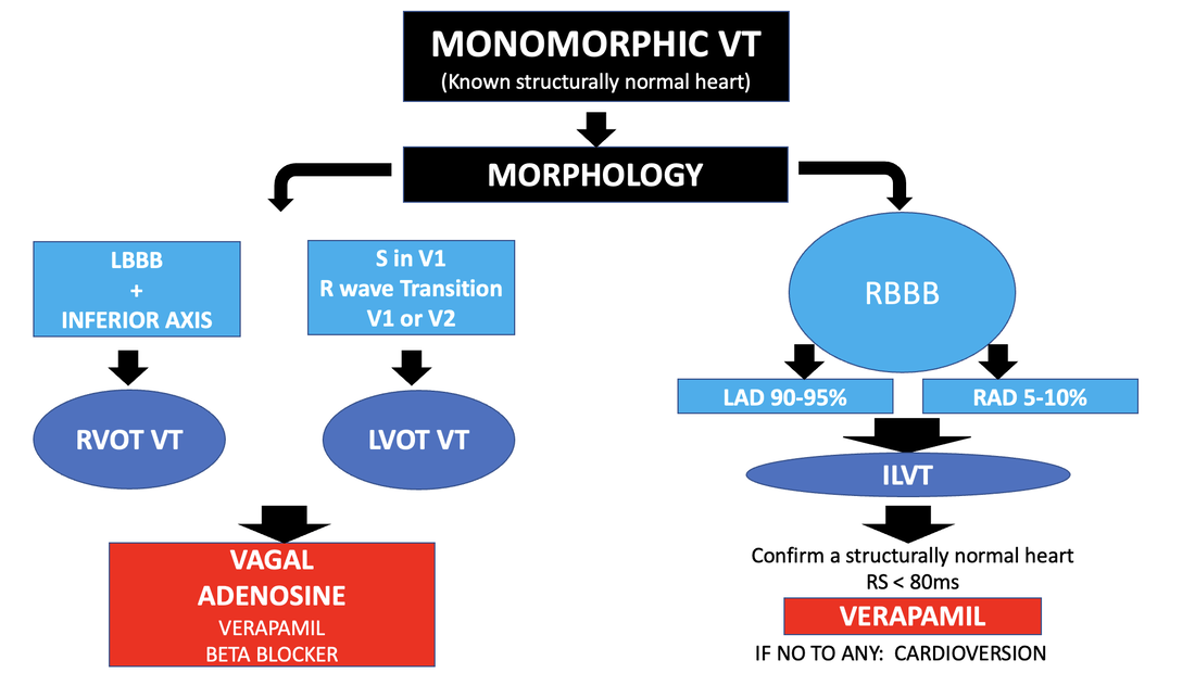
Classifications can be confusing. For simplicity we can classify them as:
  • Outflow tract VT (right or Left)
  • Idiopathic Left Ventricular tachycardia or fascicular tachycardia
​Below is a schematic on identification of each type.
Picture
1 Comment

A quiz on wide complex tachycardia

5/8/2022

0 Comments

 
I recently gave a webinar on wide complex tachycardias(WCT) and other ECGs. Here are 4 WCT ECGs for you to try. The question I would like you to answer is simple:
Is this Ventricular Tachycardia (VT)?
​Then watch the 4 minute video with the answers, below.
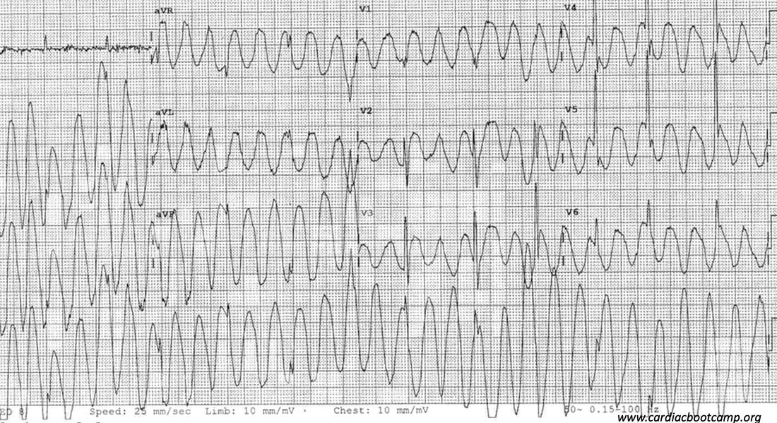
ECG 1
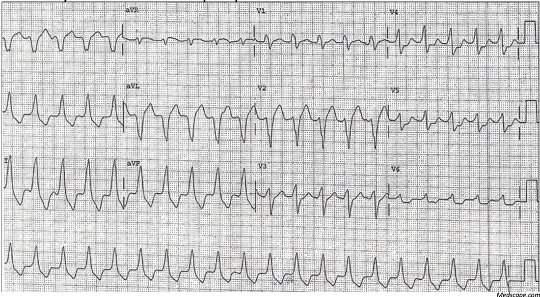
An elderly patient who is hypotensive and has had a collapse/fall is found to have a WCT on the monitor. This ECG is done. Is this VT?
Picture
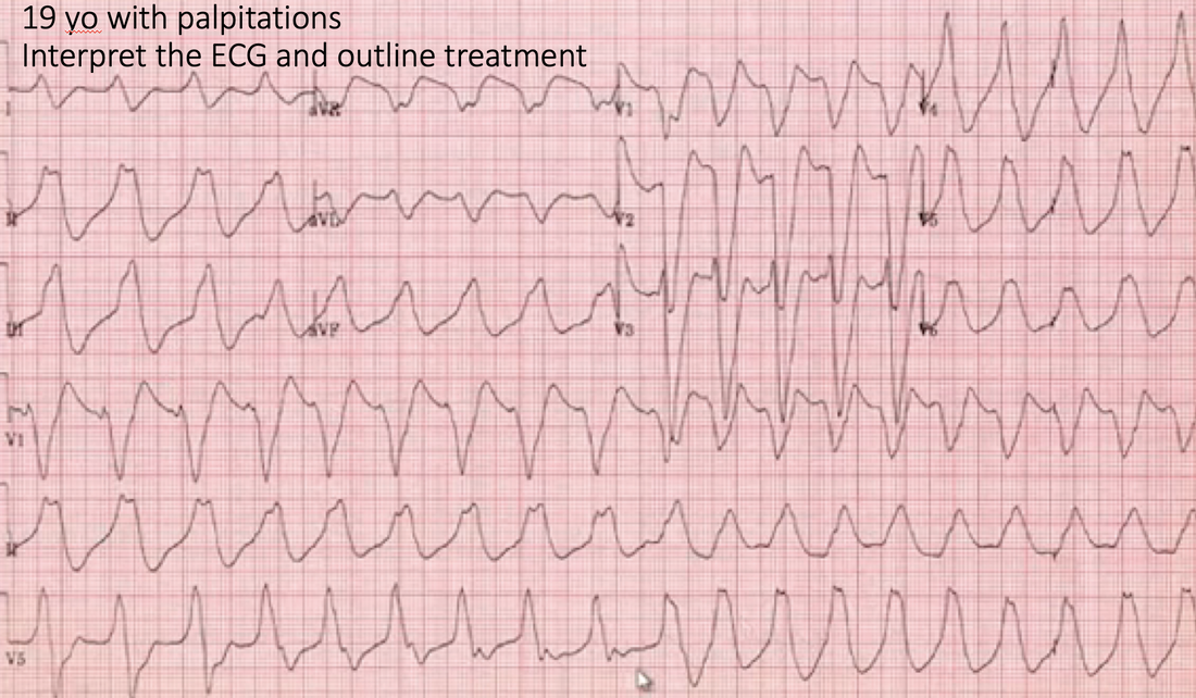
ECG 2
A 19 yo woman presents with palpitations. An ECG is done which shows this WCT. Is this VT?
Picture
ECG 3
A 58 yo patient present to the Emergency Department complaining of palpitations. Is this VT?
Picture
ECG 4
A 60 yo male presents with palpitations. Is this VT?
Picture
THE ANSWERS
0 Comments

    Author

    Dr Peter Kas
    ​Emergency Physician

    Back to resus.com.au
    Picture
    Picture
    Sign up for
    ECG of the week
     Its free

    Archives

    May 2023
    March 2023
    February 2023
    January 2023
    December 2022
    November 2022
    October 2022
    September 2022
    August 2022
    July 2022
    June 2022
    March 2022
    December 2021
    November 2021
    October 2021
    September 2021
    August 2021
    July 2021
    June 2021
    May 2021
    April 2021
    March 2021
    February 2021
    January 2021
    December 2020
    November 2020
    September 2020
    August 2020
    July 2020

    Categories

    All
    Ablation
    Atrial Fibrillation
    Atrial Flutter
    Atrial Tachycardia
    Axis
    Bradycardia
    Cardiac Arrest
    Chest Pain
    ECG Mimics
    Ischaemia
    Lead Reversal
    Migraine
    Myopericarditis
    Pacing
    Paediatric Arrhythmias
    Paediatric ECG
    Paediatric Syncope
    Pericarditis
    QT Interval
    STEMI
    STEMI Mimics
    SVT
    T Waves
    Ventricular Tachycardia
    Wellens
    Wide Complex Tachycardia

Our Other Websites to Visit

www.emcore.com.au
www.resus.com.au

Contact Us
© COPYRIGHT 2015. ALL RIGHTS RESERVED.
  • HOME
  • Course Login
    • Log In
    • Log Out
  • SYLLABUS
  • STORE
    • Online Self Study Course
    • Cardiac Video Course
  • ECG OF THE WEEK