CARDIAC BOOTCAMP
  • HOME
  • Course Login
    • Log In
    • Log Out
  • SYLLABUS
  • STORE
    • Online Self Study Course
    • Cardiac Video Course
  • ECG OF THE WEEK

ECG OF THE WEEK

BECOME A COURSE MEMBER
Each Week we put up one ECG case for you....because it's easier to learn from cases.
The ECG of the week is FREE to everyone. To become an expert at ECG's join the Cardiac Bootcamp Course. It's a great way to Learn!
​Or come to one of our LIVE Cardiac Bootcamps

48 yo with palpitations

30/5/2021

3 Comments

 
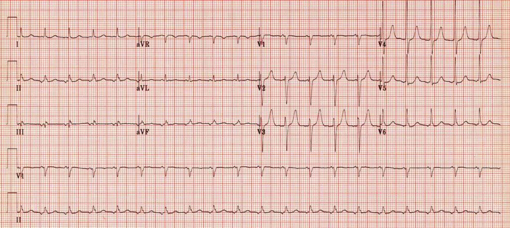
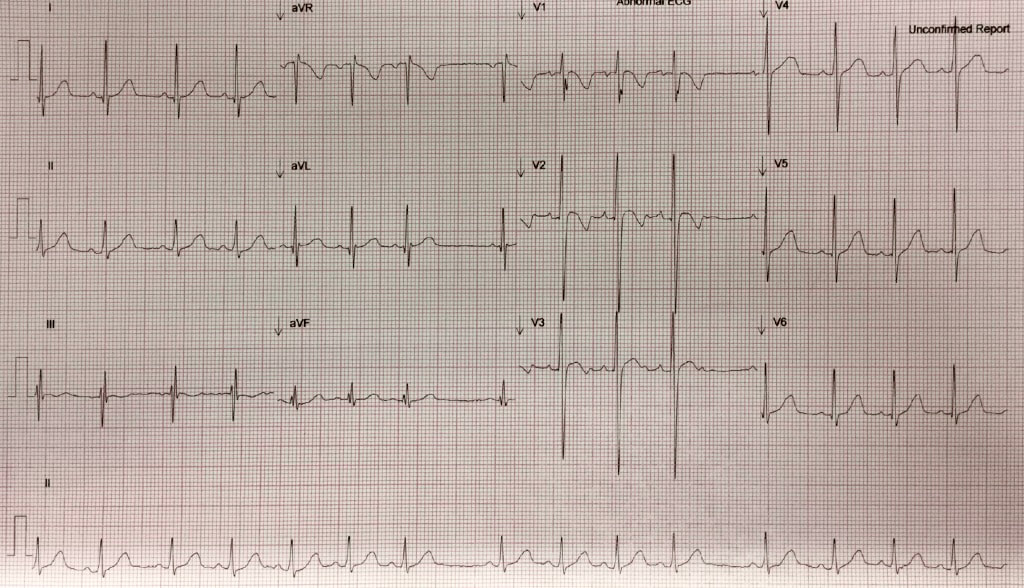
A 48 yo male presents to the emergency department with a complaint of a rapid heart rate. He has been known to be in a rapid rate before(although he is not quite sure of what it is) and has been commenced on Sotalol. He sometimes neglects to take his medication and in this case has not taken it for two days. He has also been drinking heavily the night before. He states that at 7.15 that morning he felt his heart start to race suddenly.
His ECG is shown below.
​What is your diagnosis?
Picture
In looking at the diagnosis look at the p-wave axis.
What does the ECG show?
​What is your management?
MEMBERS CLICK HERE FOR MORE
3 Comments

A 40 yo with depressed conscious state

25/5/2021

2 Comments

 
A 40 yo is brought to the emergency department with the following vitals:
  • GCS 4
  • SBP 70mmHg
  • HR 140
  • RR 50
  • T 39C
There are no signs of injury. Heart sounds are dual and no extra sounds.  There are reduced breath sounds and crepitations in the left base. The abdomen is soft and the patient appears to have peripheral cyanosis. The patient has a known history of IVDU and has been found in a chair, not rousable.
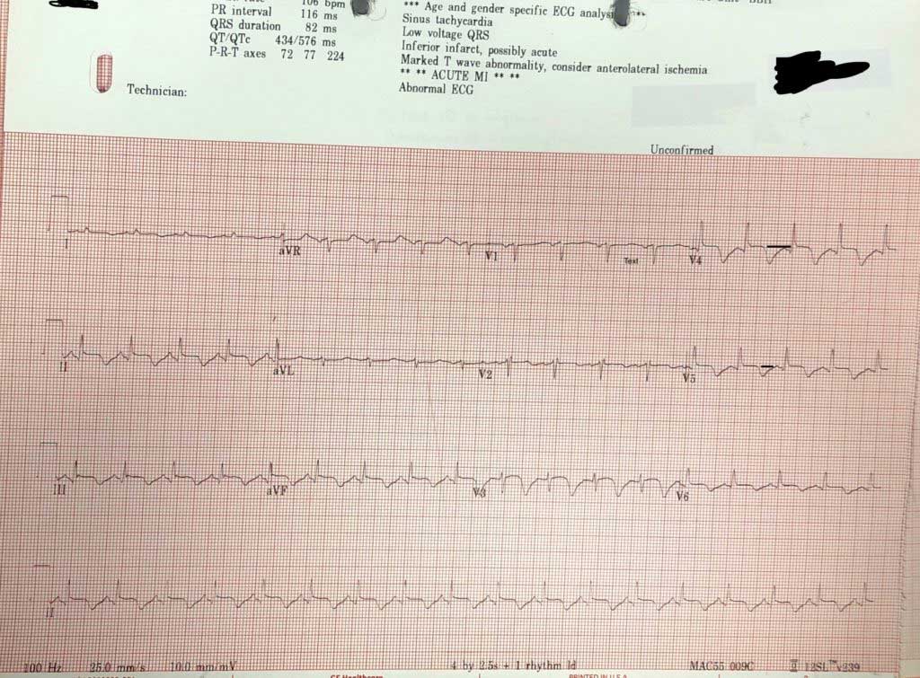
An  ECG is done.

  1. What does the ECG show?
  2. What are your differential diagnoses for this patient?
  3. What is the most likely diagnosis? 
  4. The computer says acute AMI. Is it?​​
Picture
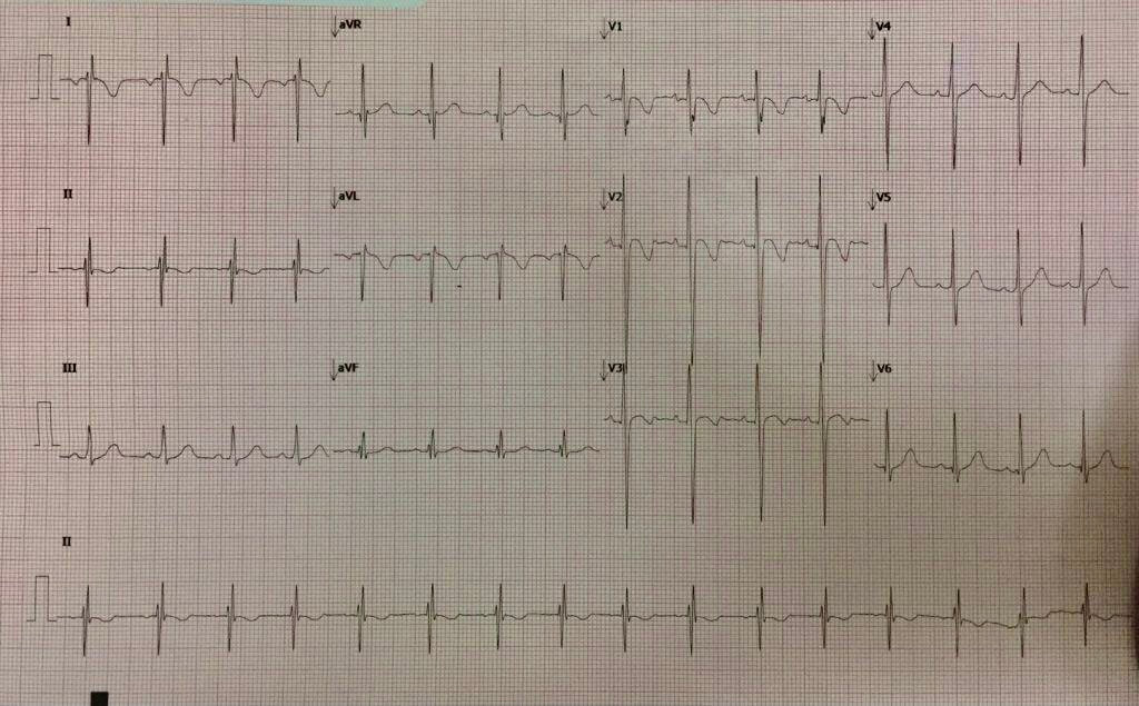
If we use the 'ECG in 20 Seconds Approach' to describe the ECG, we get the following:
Rate/Rhythm 
106

  • Is it sinus?– P waves are upright in II and inverted in aVR; Yes
  • Is there a P for every QRS? Yes
QRS:
  • Is it tall/small?: It’s about right, there is no hypertrophy.
  • Is it wide/narrow? It’s narrow
  • Is it of abnormal morphology ie., delta wave? No
  • Is it clumped?(just incase we miss a Mobitz- but it’s not slow enough) No
ST-T
  • Remember the baseline is the T-P line
    • If we look we see that there is some ST elevation in some areas of the ECG of about 1mm, although this is difficult to gauge as the T wave goes straight into the p wave.
  • The most visible abnormality is the inverted T waves.
What causes inverted T waves?
MEMBERS Click here to read the more
2 Comments

AXIS Calculation and CONGEnital heart disease in children

19/5/2021

0 Comments

 
A quick clinical pearl for you.
One of the important ECG skills to have, is to be able to diagnose a paediatric ECG. Go to the Paediatric ECG page in the course to quickly read on this.

There are the important things to know which include:
  • Kids are right heart dominant, then progressively become left heart dominant
  • The intervals are a little different
  • The T waves are upright in the first week of life and then flip until adolescence
................ etc

What about the axis?
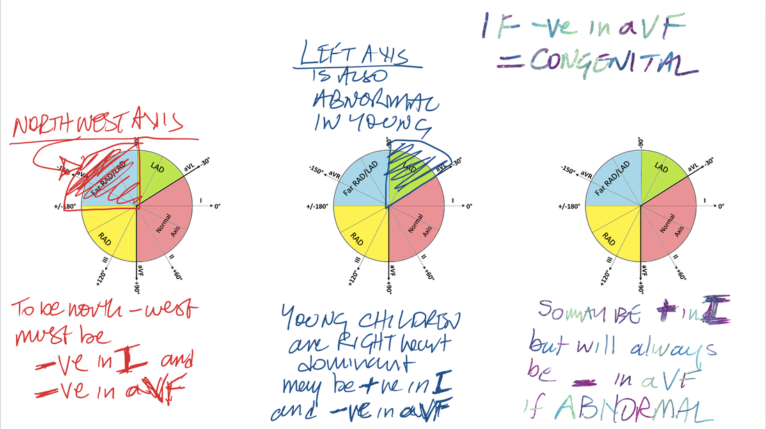
One of the first things I look at is the AXIS. The paediatric axis should be normal or rightward. If the axis is NORTHWEST, assume a congenital abnormality like a VSD etc.

JUST LOOK at aVF.  If aVF is NEGATIVE= Congenital until proven otherwise.
Picture
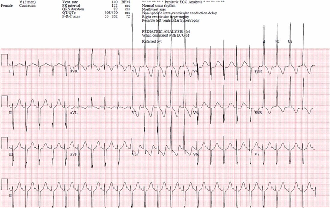
Below is an example of this. It is a child with an AV Septal Defect, causing biventricular failure. The ECG should be right heart dominant, but it's both right and left dominant.
Look at aVF; it's NEGATIVE. So I know there is a congenital defect.
How will these children present. What are the clinical manifestations of this conditions and others? Go to Paediatric Heart Conditions you must know.
Picture
Source: Pediatrics in Review
0 Comments

A 9 yo girl with collapse

10/5/2021

1 Comment

 
A 9 yo girl is brought into the emergency department, following an episode of syncope. She has a current viral illness and looks miserable. She was walking into the Emergency Department with her mother, when she collapsed. She recovered quickly with no post-ictal component but has no memory of the event, although the event was preceded by ‘dizziness’ and diaphoresis.  Her past medical history is ADHD, for which she is on no medication.
You review her in the triage room as the department is filled to capacity. The vitals are all normal but the child looks tired and has some coryza symptoms. You ask the resident to get the rest of the story and show you an ECG.
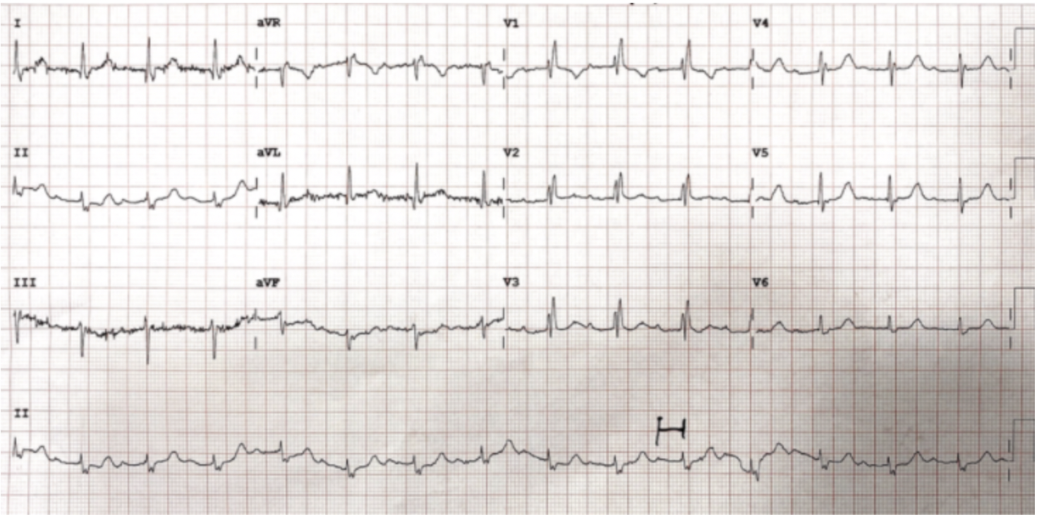
The resident brings you the ECG shown below. What is your comment on this ECG?
Picture
ECG Interpretation
At first glance: There is T wave inversion in anterior leads but you would expect this in a young girl. There are however T waves inverted in the lateral leads…….STOP. Let’s go over the ECG in a systematic fashion.
Is it sinus rhythm? For it to be sinus, the p wave in II must be upright and the p wave in aVR inverted. This is not the case. Now look at lead I, The p waves are inverted and they should be upright.
Two things give you this pattern:
  1. Limb lead reversal and
  2. Dextrocardia
This ECG is the result of limb lead reversal.
You have the ECG redone. The ECG is shown below. How would you comment on this?
Picture
Cardiac Bootcamp Self Study Course Members
should read the section on Paediatric ECG

Read More
1 Comment

IS calculating axis of any use?

6/5/2021

0 Comments

 
For quite some time I've been less than eager to calculate axis on all ECGs. The one exception that I made is, when there was a RBBB on the ECG, as I would then look for a Fascicular Block.
However I have found it more useful than I thought.
In this Blog we will look at how to calculate axis and refer you back to the section in the course on ECG Axis to do this in more detail and we will further look at some of the uses.
There are many ways to calculate axis;
  • the quadrant method,
  • the 3 lead method,
  • the degree method and more.
The difference is in the preciseness with which you wish to calculate the axis ie., are you happy to say it's normal or it's leftward, or do you need to know that this axis is not only rightward, but that it is 95 degrees.
WHY IS IT IMPORTANT?
There was a case we covered in a prior blog of a 70 yo male who presents to the emergency department following a collapse at home. There is no prodrome and he does not remember the incident.
​His ECG is shown below. What is the diagnosis?
Picture
This was an incomplete triphasicular block, with:
  • RBBB
  • First degree block
  • left axis ie LAFB
Read the section on fascicular blocks to find out more.
The next case is one of an amphetamine overdose. Can the axis help us here?
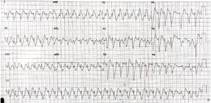
​The patient has been in a sinus tachycardia and now he has a wide complex.
  • Is it VT?
  • Is it Na channel Blockade?
  • Is it RBBB?
Picture
It is a wide complex tachycardia. It has a rate > 120, so it qualifies to continue with my 120CRAM formula for diagnosing VT. It has some of the elements of VT.
First thing to say is that amphetamines DO NOT cause Na channel blockade, so it can't be that.
What is the axis? It looks like a right axis. Right axis can be cause by ventricular ectopy ie VT. So it can be VT.
RBBB doesn't tend to cause a right axis. BUT HyperK+ does and this patient was hyperkalaemic. After being shocked with no result, the patient was treated with bicarb and Ca++ and sinus rhythm was obtained.
Axis can help.
HOW TO QUICKLY CALCULATE AXIS: Let's use the Quadrant Method
We will use leads I and aVF
Here is how to perform this:
  • Assign Lead I to your LEFT ARM
  • Assign Lead aVF to your RIGHT ARM
Now look at the leads and raise the arm belonging to the positive lead and lower the arm belonging to the negative lead. The arm left upward will be the axis if Left or right.
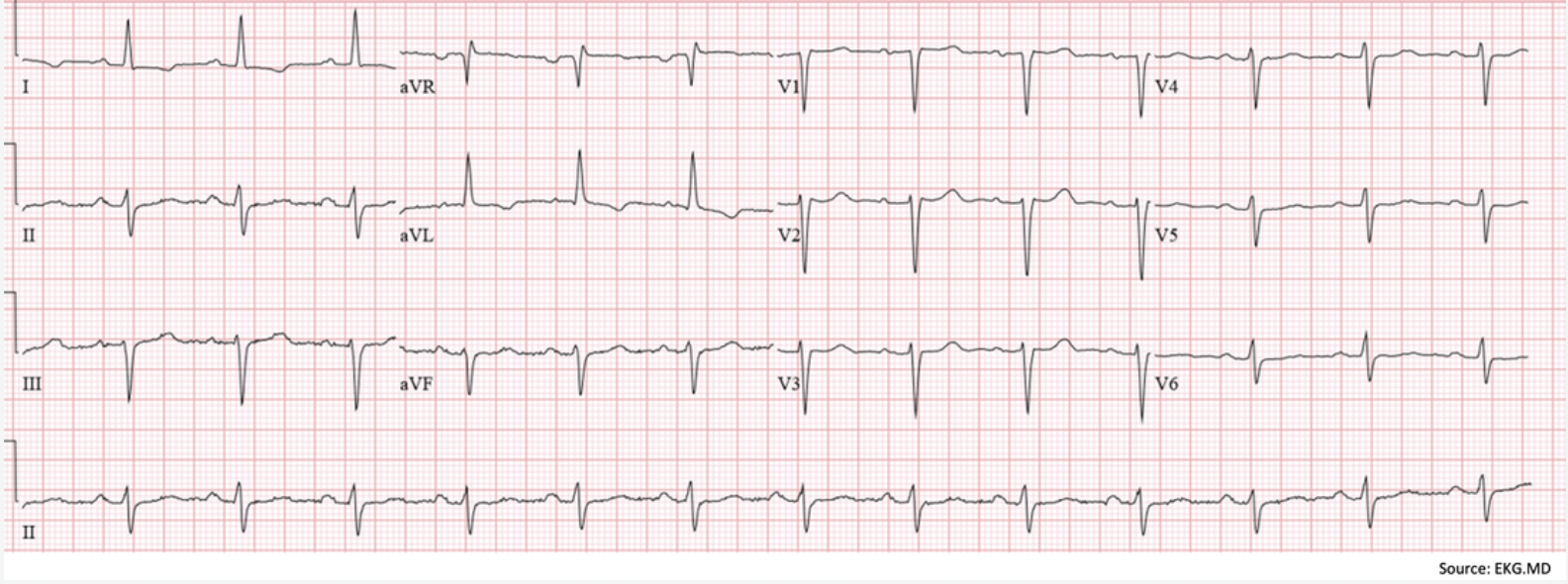
Let's practice on the ECG below:
Using the Quadrant Method the left arm goes up as lead I is positive and the right arm is downas aVF is negative. The resultant positive arm is the left so it is a LEFT AXIS. 

Picture
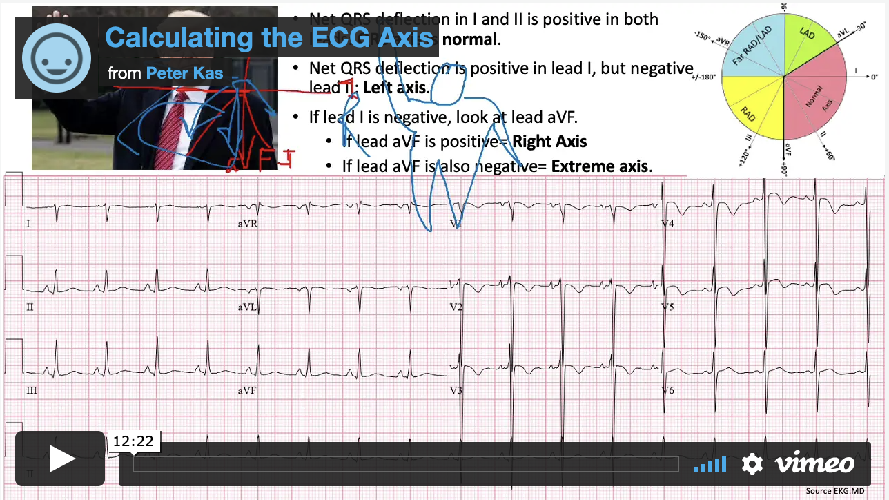
For a Video on how to do this and a bit of fun with Donald Trump showing us how to calculate axis, go to the Basic ECG Section.
Picture
That's all I've got for you this week....
0 Comments

    Author

    Dr Peter Kas
    ​Emergency Physician

    Picture
    Picture
    Sign up for
    ECG of the week
     Its free

    Archives

    February 2023
    January 2023
    December 2022
    November 2022
    October 2022
    September 2022
    August 2022
    July 2022
    June 2022
    March 2022
    December 2021
    November 2021
    October 2021
    September 2021
    August 2021
    July 2021
    June 2021
    May 2021
    April 2021
    March 2021
    February 2021
    January 2021
    December 2020
    November 2020
    September 2020
    August 2020
    July 2020

    Categories

    All
    Atrial Fibrillation
    Atrial Flutter
    Atrial Tachycardia
    Axis
    Bradycardia
    Cardiac Arrest
    Chest Pain
    ECG Mimics
    Ischaemia
    Lead Reversal
    Myopericarditis
    Pacing
    Paediatric Arrhythmias
    Paediatric ECG
    Paediatric Syncope
    Pericarditis
    QT Interval
    STEMI
    STEMI Mimics
    SVT
    Ventricular Tachycardia
    Wellens
    Wide Complex Tachycardia

Our Other Websites to Visit

www.emcore.com.au
www.resus.com.au

Contact Us
© COPYRIGHT 2015. ALL RIGHTS RESERVED.
  • HOME
  • Course Login
    • Log In
    • Log Out
  • SYLLABUS
  • STORE
    • Online Self Study Course
    • Cardiac Video Course
  • ECG OF THE WEEK