CARDIAC BOOTCAMP
  • HOME
  • Course Login
    • Log In
    • Log Out
  • SYLLABUS
  • STORE
    • Online Self Study Course
    • Cardiac Video Course
  • ECG OF THE WEEK

ECG OF THE WEEK

BECOME A COURSE MEMBER
Each Week we put up one ECG case for you....because it's easier to learn from cases.
The ECG of the week is FREE to everyone. To become an expert at ECG's join the Cardiac Bootcamp Course. It's a great way to Learn!
​Or come to one of our LIVE Cardiac Bootcamps

The Non-Specific T wave abnormality

16/5/2023

1 Comment

 
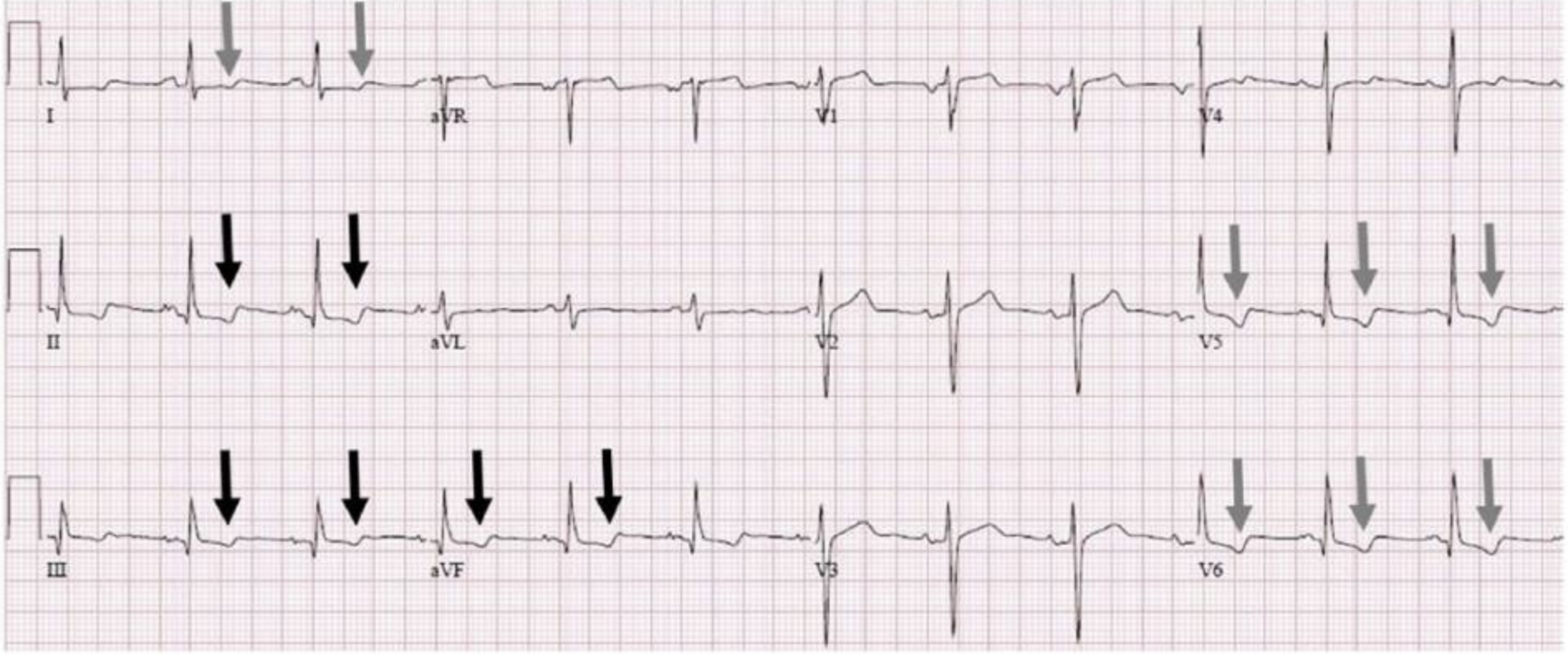
A 72 yo male patient presents with chest pain. The pain is sharp and is worst on lying down. There is a past history of hypertension, high cholesterol and a family history of heart disease. ​An ECG is done and shown below (1).
Picture
T wave inversion in the Inferior and Lateral leads
The ECG is normal except for some non-specific T wave changes in the inferior and lateral leads. What do we do with these changes? Is this inferolateral ischaemia? This patient has ECG changes so needs to be worked up as per a chest pain stratification and pathway tool. Is there anything more that we can do to work out if these changes are cardiac in origin?

What are non-specific T wave changes?
They occur in about 1% of patients and  include T wave flattening and T wave inversion with no other changes necessarily present. 
Causes include(1):
  • Myocardial Infarction
  • Myocarditis
  • Mitral valve prolapse
  • Ventricular Strain.
Can changes be due to respiration?
Respiration results in a change in heart position. The heart rotates in a clockwise direction  in inspiration and anticlockwise in expiration. This can cause T wave changes. Inspiration also produces diaphragmatic movement, which can cause T wave changes (2)
A 2017 case report(1) looked at one case where non-specific T wave changes had a respiratory cause and recommends we look for this.
The authors recommend that:
  1. An ECG be performed with the breath held in deep inspiration and
  2. A second ECG be performed with the breath held in in expiration.
A change in the polarity of the T waves with respiration may indicate a non-cardiac cause is more likely.

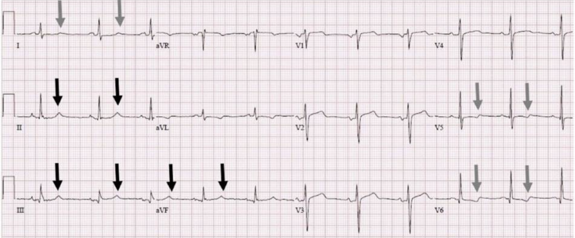
The ECG below was done in inspiration.
Picture
In this ECG, the patient holds their breath at the end of inspiration.
In the above ECG, the T waves in the inferior leads are now upright. The T wave morphology has also changed in the lateral leads.
This indicates a potential respiratory cause.
We must beware however, as this patient has risk factors and is not really low risk:
  1. If we applied a Heart Score to this patient, it would be 5 even if the troponin were normal, so a moderate risk.
  2. If EDACS was applied, again they are not a low risk patient.
This patient would still benefit from serial troponins and being assessed on a chest pain pathway.
​
References
  1. Sharma H and Tiwari A. Respiratory T-Wave inversion in a Patient With Chest Pain. Clinical Medicine Insights: Case reports. 2017;10: 1-3
  2. Dougherty JD. The relation of respiratory changes in the horizontal QRS and T-wave axes to movement of the thoracic electrodes. J Electrocardiol. 1970;3:77–86.
1 Comment

HYPERACUTE T WAVES: STEMI EQUIVALENT

23/3/2023

0 Comments

 
A 60 yo patient presents with chest pain. He has a past medical history of hypertension and high cholesterol and has diet-controlled diabetes.
He is haemodynamically stable, with a normal clinical examination.
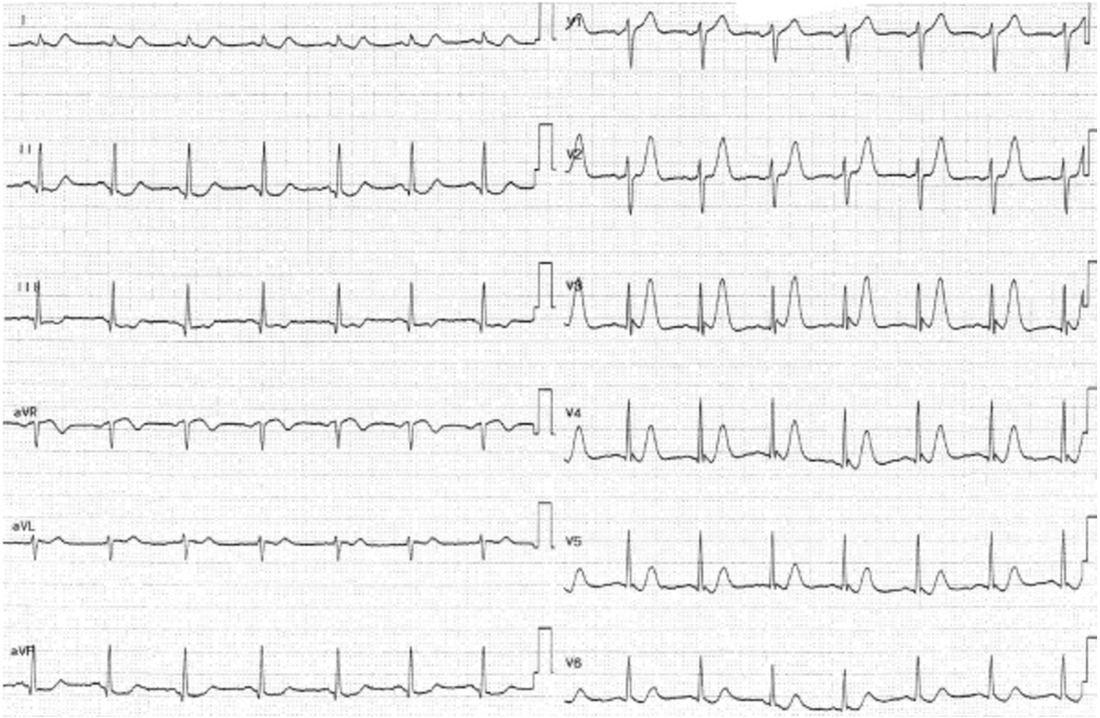
His ECG is shown below.
Picture
There are definitely hyper acute T waves. What does that actually mean? It's important to note that here is no formal definition of a hyper acute T wave. However it is not based on amplitude alone.
The hyper acute T wave is considered a STEMI-equivalent.

Koechlin L et al(1) conducted a post hoc analysis  of a multicenter diagnostic study and looked at the diagnostic performance of T wave amplitude for diagnosing myocardial infarction. They found that they were not useful in making this diagnosis.

One of the concerns is that they only looked at T wave amplitude. Smith(2) states that more than just amplitude is important to make the diagnosis. He refers to the T-wave 'bulk' The T wave is large in area, symmetric relative to the QRS.

The term " T wave towers" over the R wave in V3, or even that the QRS can fit into the T wave are important in terms of this 'bulk'.

References
  1. Koechlin L et al. Hyperacute T Wave in the Early Diagnosis of Acute Myocardial Infarction. Ann of Emerg Med.2022; 1-9
  2. Smit S et al. Hyperacute T-waves Can be a Useful Sign of Occlusion Myocardial Infarction if Appropriately Defined. Annals of Emerg Med. 2023;1-4
0 Comments

    Author

    Dr Peter Kas
    ​Emergency Physician

    Back to resus.com.au
    Picture
    Picture
    Sign up for
    ECG of the week
     Its free

    Archives

    May 2023
    March 2023
    February 2023
    January 2023
    December 2022
    November 2022
    October 2022
    September 2022
    August 2022
    July 2022
    June 2022
    March 2022
    December 2021
    November 2021
    October 2021
    September 2021
    August 2021
    July 2021
    June 2021
    May 2021
    April 2021
    March 2021
    February 2021
    January 2021
    December 2020
    November 2020
    September 2020
    August 2020
    July 2020

    Categories

    All
    Ablation
    Atrial Fibrillation
    Atrial Flutter
    Atrial Tachycardia
    Axis
    Bradycardia
    Cardiac Arrest
    Chest Pain
    ECG Mimics
    Ischaemia
    Lead Reversal
    Migraine
    Myopericarditis
    Pacing
    Paediatric Arrhythmias
    Paediatric ECG
    Paediatric Syncope
    Pericarditis
    QT Interval
    STEMI
    STEMI Mimics
    SVT
    T Waves
    Ventricular Tachycardia
    Wellens
    Wide Complex Tachycardia

Our Other Websites to Visit

www.emcore.com.au
www.resus.com.au

Contact Us
© COPYRIGHT 2015. ALL RIGHTS RESERVED.
  • HOME
  • Course Login
    • Log In
    • Log Out
  • SYLLABUS
  • STORE
    • Online Self Study Course
    • Cardiac Video Course
  • ECG OF THE WEEK