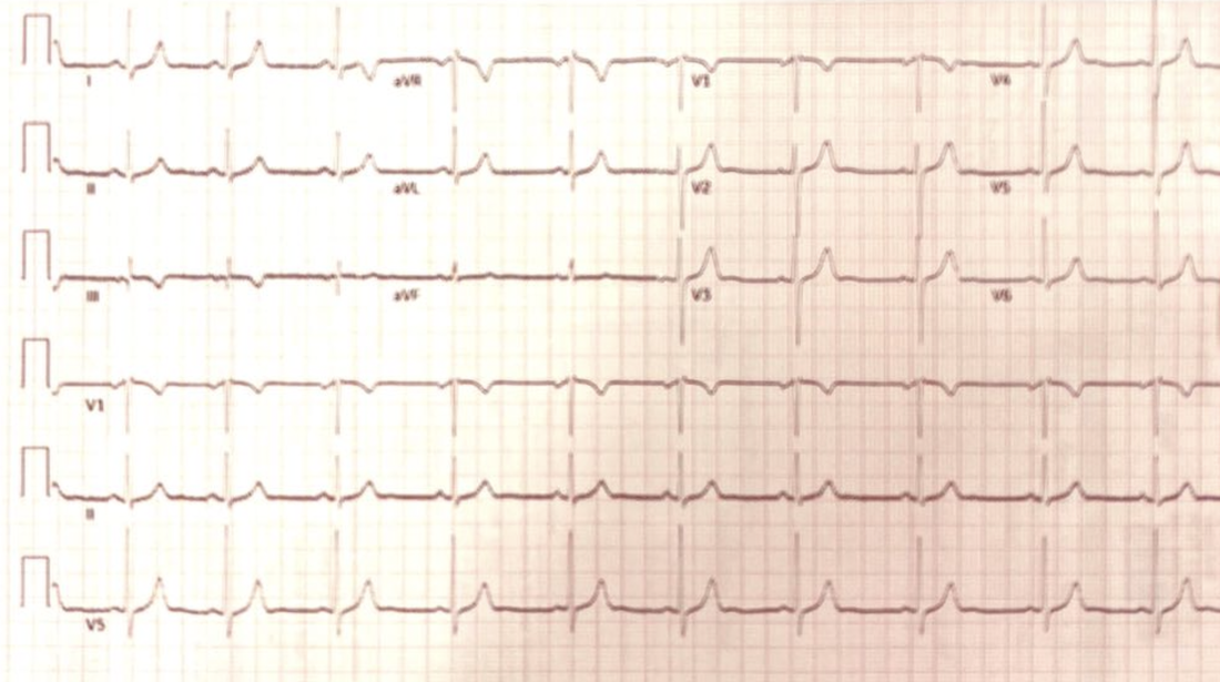
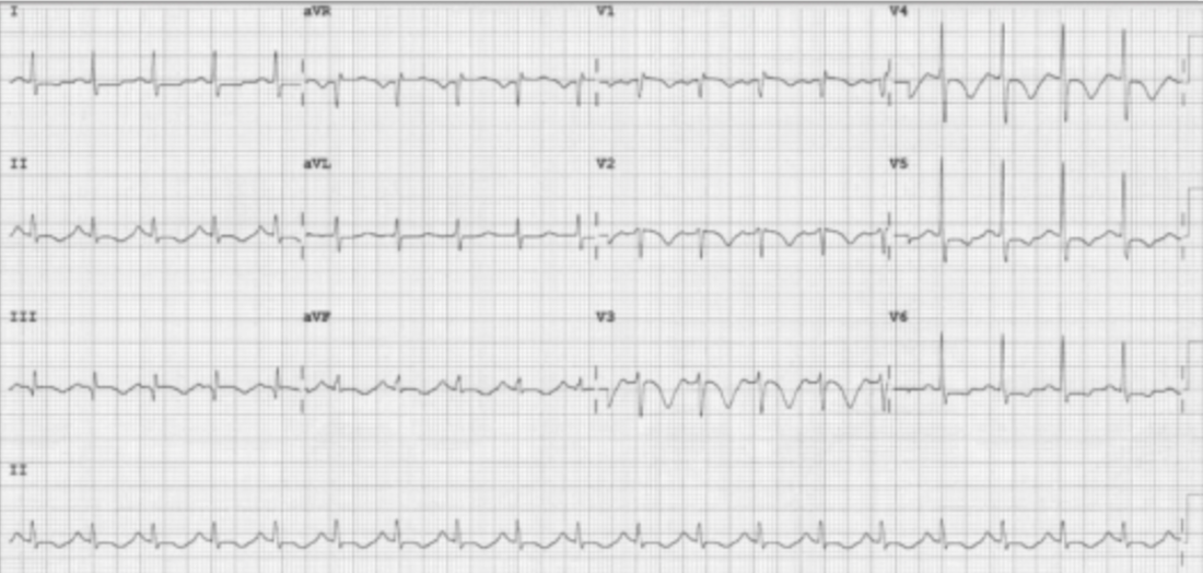
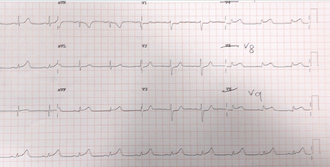
CARDIAC BOOTCAMP
  • HOME
  • Course Login
    • Log In
    • Log Out
  • SYLLABUS
  • STORE
    • Online Self Study Course
    • Cardiac Video Course
  • ECG OF THE WEEK

ECG OF THE WEEK

BECOME A COURSE MEMBER
Each Week we put up one ECG case for you....because it's easier to learn from cases.
The ECG of the week is FREE to everyone. To become an expert at ECG's join the Cardiac Bootcamp Course. It's a great way to Learn!
​Or come to one of our LIVE Cardiac Bootcamps

a 48 yo male with palpitations

14/12/2021

0 Comments

 
A 48 yo male presents to the emergency department with a complaint of a rapid heart rate. He has been known to be in this before and is usually on Sotalol, but has not taken any for two days. He has also been drinking heavily the night before. He states that at 7.15 that morning he felt his heart start to race suddenly. His ECG is shown below. What is your diagnosis?
Picture
This ECG shows an atrial tachycardia with atrial rate of 120bpm. The p waves are inverted in III and aVF, which is abnormal. This is a unifocal atrial tachycardia. It can occur in structurally normal and abnormal hearts. it arises from a particular part of the atrium and its location can be diagnosed by the axis of the p waves.
Below is another example:
Picture
This is someone at a rate of 120bpm. Notice the inverted p’s in lead III. Again, this is unifocal atrial tachycardia.
Clinical Presentation
The usual presentation is one of palpitations, but may be that of syncope, or cardiac failure if the patient has been in the rhythm long enough.
Workup
An ECG and electrolytes to ensure no abnormalities and a full blood count to exclude anaemia.
Very few other investigations are needed in the emergency department. Perhaps a chest xray if lung pathology is suspected. The patient should have an echocardiogram, to look for structural abnormalities, but this can be as an outpatient.
Management
The unifocal atrial tachycardia can be treated like an SVT.
The general treatment for this condition is:
  1. Pharmacological
    1. Beta blockers
    2. Calcium channel blockers
  2. Cardioversion
In this case we gave the patient his usual dose of Sotalol and waited, however it was clear that there was not going to be resolution, so we proceeded to cardiovert.

0 Comments

the vt score: 100% specificity for vt

14/12/2021

0 Comments

 
Introduction
When we assume that every wide complex tachycardia is Ventricular Tachycardia(VT) our sensitivity for picking up VT is 100%. In the emergency department setting, this is a must! We use electricity on all of these patients because it is safe and effective. We can try adenosine, in case it’s a supra ventricular tachycardia(SVT) but only if it’s a regular rhythm.
Our specificity in this scenario lags, however in our favour, is that fact that 80% of WCT is VT. If we wanted to determine if an ECG is showing VT, we have a range of rules at our disposal,  including Brugada and Vereckei, that give a respectable specificity, but it’s still in the mid 80%.
A new validated rule
The VT Score, uses 7 criteria, some of which we already use, to diagnose VT.  A specificity of 100% is reached when 4 or more criteria are satisfied(1).
Picture
The Seven Criteria are as follows:
NOTE: Before we start I would add that the rate should be greater than 120 beats per minute for us to start considering VT.
1. Dominant Initial R wave in V1. This can be notched(the original rabbit’s ear) provided the notch is on the descending limb.
Picture
2. Initial r > 40 ms in V1 or V2: assess in predominantly negative QRS complexes. Look for a ‘fat’ initial r in rS in V1. It can also include variations such as: RS with high ‘r’ amplitude, rS with notched r
Picture
3 Notched S in V1. The notch can be at the beginning, in the middle or near the nadir of the S wave.(This has been known as Josephson’s Sign)
Picture
4. Initial R wave in aVR. The QRS in aVR must start with a large R wave. it can be notched. (This comes from the Vereckei(2) criteria).
Picture
5. Lead II R wave Peak time(RWPT) > or equal to 50ms. This is the time from the beginning of QRS to first change in direction.
Picture
6. Lack of RS in V1-V6.(Brugada Criterion(3)). This is similar to saying that there is concordance.
Picture
7. Atrioventricular Dissociation. This is indicated where a fast ventricular activity iOS not related to atrial depolarisation.
Picture
Source: LITFL
LETS MAKE IT EASIER TO REMEMBER:
  1. Rate must be > 120bpm to be VT
  2. Concordance in V1-V6
  3. AV Dissociation
  4. Josephson’s Sign in V1 -V2
  5. Look at V1 for a FAT INITIAL R wave (r>40ms) this could be the rabbits ear
  6. Look at aVR for a FAT Wave to begin with
  7. Look at lead II RWPT > 50ms- again a big R wave.
References
  1. Jastrzebski M et al. The ventricular tachycardia score: a novel approach to electrocardiographic diagnosis of ventricular tachycardia. Europace. 2016 Apr;18(4):578-84
  2. Vereckei A et al. Application of a new algorithm in the differential diagnosis of wide QRS complex tachycardia. Eur Heart J 2007.28:589-600
  3. Brugada P. et al. A new approach to the differential diagnosis of a regular tachycardia with a wide QRS complex. Circulation 1991:83:1649-1659
0 Comments

a 6 week old with poor feeding

14/12/2021

0 Comments

 
A 6 week old baby is brought into the emergency department by her mother with irritability and poor feeding. She is afebrile and her examination reveals bilateral crepitations in the chest and a systolic murmur. What does the ECG show and what is the likely diagnosis?
Picture
The ECG is usually quite straightforward to analyse if we remember 2 things:
  1. The T waves are upright in the first week of life. They are inverted after this until about age 9-10
  2. When a child is born, they are right heart dominant. As they grow older, they become more left heart dominant.
Any neonate or infant presenting with difficulty feeding, or shortness of breath whilst feeding, or change of colour whilst feeding, needs to be examined for a potential heart condition.
In any child presenting this way, we need to think of:
  1. VSD
  2. Tetralogy of Fallot
  3. Myocarditis
  4. Cardiomyopathy
  5. Arrhythmia.
It is also useful to have an idea of heart rates at various age groupings. My rough guide is:
  • < 2yo max  160
  • 2 – 7yo max 140
  • 7 – 15 yo max 130
Here is the approach as per the ‘ECG in 20 Seconds’
Rate: just count the number of complexes and multiply by 6 ( an ECG takes 10 seconds to print). Rate = 23 x 6 = 138, which is acceptable
Rhythm: For sinus rhythm, there must be a P wave before each QRS and the P wave must be upright in II and inverted in aVR. If not, then it is not sinus rhythm and the pacemaker is in the low right/left atrium
This is sinus rhythm 
QRS: is it too tall or too small, ie is there hypertrophy? Remember that in the child the is right ventricle is dominant. To judge hypertrophy look at V6. If the R wave intersects the baseline of the V5 tracing above, then there is LVH
In terms of Right Ventricular Hypertrophy, in adults it is usually hidden by the left ventricle, however in children the clues are in  V1. There is RVH if there is an rsR pattern in V1, or a pure R wave in V1 after 6 months.
It is also helpful to look at the R wave to S wave ratio. Remember that the young heart is right heart dominant, so you would expect that the R wave is more predominant than the S wave in the right leads, however if the opposite is true, then there is left ventricular hypertrophy.
The QRS is normally +ve in aVF
  • If it is negative in aVF, there may be cardiac malformations, such as AV septal defect.
  • If it is biphasic in aVF, it may be normal, but needs cardiology discussion.
Look for the abnormal QRS morphology i.e.., the delta wave of WPW
This shows Left Ventricular Hypertrophy. The heart is left heart dominant rather than right heart dominant, which we would expect in this age group. With the murmur and the crepitations and the left heart dominance, this is most likely VSD(left to right shunt).
ST-T: The T wave is upright for the first week of life, then is inverted until adolescence.
This is normal.
Intervals:
PR- In anyone, an interval > 0.2 sec is abnormal
  • In infants and young children an interval of > 0.16 (4 small squares) is abnormal.
QT- >0.45sec is abnormal in anyone
  • In < 6 months <0.49sec
  • In > 6 months <0.44
It appears that the QTc is quite long here.
This ECG is of a patient with VSD. Paeds referral is needed for further investigation including an Echo and bloods. Treatment may include:
  • Oxygen
  • Fluid Restriction
  • Diuretics
  • Ace Inhibitor
0 Comments

40 yo with a depressed conscious state

14/12/2021

1 Comment

 
​The Case
​A 40 yo is brought to the emergency department with the following vitals:
  • GCS 4
  • SBP 70mmHg
  • HR 140
  • RR 50
  • T 39C
There are no signs of injury. Heart sounds are dual and no extra sounds.  There are reduced breath sounds and crepitations in the left base. The abdomen is soft and the patient appears to have peripheral cyanosis.
The patient has a known history of IVDU and has been found in a chair, not rousable.
She has fluids and inotropes and antibiotics and she has the ECG shown.
What are your differential diagnoses for this patient? What is the most likely diagnosis? 
The computer says acute AMI. Could it be?
Let’s look at the ECG
Picture
Rate 106
Is it sinus?– P waves are upright in II and inverted in aVR; Yes
Is there a P for every QRS? Yes
QRS:
Is it tall/small?: It’s about right, there is no hypertrophy.
Is it wide/narrow? It’s narrow
Is it of abnormal morphology ie., delta wave? No
Is it clumped?(just incase we miss a Mobitz- but it’s not slow enough) No
ST-T
Remember the baseline is the T-P line
If we look we see that there is some ST elevation in some areas of the ECG of about 1mm, although this is difficult to gauge as the T wave goes straight into the p wave.
The most visible abnormality is the inverted T waves.
What causes inverted T waves?As this is an adult patient we can overlook the T wave inversion of childhood. Other prominent causes are:
  • Acute Myocardial Infarction
  • Ventricular Hypertrophy( not present on this ECG)
  • Pulmonary Embolism
  • Raised Intracranial Pressure( I would usually expect them to be deeper)
PR and QT Intervals
The PR is normal and the QT is very prolonged.
PACING Spikes: None.
Putting it all together, what would seem the most probable diagnosis?Before we look at the most likely diagnoses. One differential to entertain is an overdose, however there are no signs of classic QRS widening of Na channel blockade, or a terminal R wave in aVR.
It might be an AMI, there is no evidence of hypertrophy, the patient is not behaving like they have a PE, but they may have raised intracranial pressure given the decreased conscious state. The decreased conscious state may be simply due to low blood pressure.
The patient is also febrile. Why? Remember this patient has a history of IVDU, so infective endocarditis is a consideration. The patient may also have meningitis. The fever may also be secondary to stroke as we know that about 50% of stroke develop a fever, which in itself is a marker of severity and marker of mortality.
Let’s narrow it down to 2 causes, AMI and raised intracranial pressure. Raised intracranial pressure ECG’s have deep T wave inversion and a prolonged QT. I’m thinking this is an intracranial event, especially if the fever is due to this.
Echo showed a myocardium, not contraction properly, with clean valves, so it may be AMI. However the CT gave the diagnosis with large subarachnoid haemorrhage.
This is a neurocardiogenic phenomenon when the heart is affected by deranged autonomic supply coming from a stroke. A fascinating case mixed with electrolyte abnormalities etc. I hope you enjoyed it.
1 Comment

31 yo female, G1P0 with pleuritic chest pain

8/12/2021

0 Comments

 
A 31 yo female presents to the emergency department with pleuritic chest pain. The patient is 19/40 gestation and no medical conditions. The history of the event is that the patient was sitting on the couch and developed left shoulder and then left chest pleuritic chest pain. There was no history of trauma, or fevers, cough or wheeze and the patient had no history of a DVT.
Clinical examination was normal.
Basic blood tests were done that showed:
CRP 4
D-Dimer 0.71 mg/L
Na 134/ K 4.1/ HCO3 19/ Creat 61/ eGFR 90
WCC 10.4/ Neut 6.3/
High sensitivity troponin 3
Vitals were HR 80bpm, BP 122/70 SpO2 100% on RA.
​What is your working diagnosis so far?
​An ECG was done as shown below. The resident reported it as normal. What does it show and what is your diagnosis?
Picture
ANSWER
This is a normal sinus rhythm ECG, with good R wave progression. The only finding of note is S1Q3T3
Take Part in the Poll

Who is your favorite singer?

:
DAYS
:
HRS
:
MINS
SECS

POWERD BY BOOMTECH
x x
What are some of the important ECG changes seen in Pulmonary Embolism?
  • S1Q3T3- +ve LR 3.7 of a PE(1), although often said to be neither sensitive nor specific.
  • RBBB
  • T wave inversion V1-V4 +ve LR 3.7
  • Tachycardia +ve LR 1.8
What did the YEARS Study tell us?
In 2017, we were given a new approach to looking at PE and the D-dimer(2)
They used a clinical decision rule which consisted of 3 items:
  1. Clinical Signs of Deep Venous Thrombosis
  2. Haemoptysis
  3. Is Pulmonary Embolism the Most Likely Diagnsosis?
The flow diagram is shown below, but in summary it says:
In the non-pregnant patient:
  • If no criteria can rule out PE with d-dimer of <1000 ng/mL
  • If criteria can rule out PE with a D-dimer of <500ng/mL
In the pregnant patient:
  • If no criteria can rule out PE if D-dimer is < 1000 ng/mL 
  • If criteria can rule out PE if D-dimer is <500 ng/mL
OVERALL
  • In any patient with NO CRITERIA a D-dimer of <1000 ruled out PE
  • In any patient WITH CRITERIA a D-dimer of < 500 rule out PE
Picture
(2)
The Pregnancy Adapted algorithm from 2019(3) is shown below:
Picture

WHAT HAPPENED TO THE PATIENT?

It was decided that although pleuritic chest pain was all that the patient had, there was nothing else to explain the symptom. The D-dimer was > 500, so the patient was sent for a CTPA. It was negative. The patient was discharged for GP followup.
Members, go to the section on PE in the course and on the ECG's of PE to see more.

References
  1. Marchick M.R. et al. 12 Lead ECG Findings of Pulmonary Hypertension occur more frequently in Emergency Department patients with Pulmonary Embolism than in patients without Pulmonary Embolism. Annals of Emergency Medicine. Vol 55, Issue 4, April 01, 2010; pp 331-335
  2. Van der Hulle T et al. Simplified diagnostic management of suspected pulmonary embolism (the YEARS study): a prospective, multicentre, cohort study. Lancet. 2017; 390:289-297.
  3. Van der Pol L.M et al. Pregnancy-Adapted YEARS Algorithm for Diagnosis of Suspected Pulmonary Embolism.N Engl J Med 2019; 380:1139-1149
0 Comments

IT'S TIME FOR A QUICK ECG QUIZ!

2/12/2021

1 Comment

 
This week I have 4 questions for you. 3 are ECGs you must know and the fourth is a simple question on what you should look for in the QRS complex, when reading the ECG. Enjoy. Hope to see you at the Live Cardiac Bootcamp Course on the Gold Coast in January 2022. Congratulations to everyone who has purchased the Online Cardiac Course and thank you for all the great comments. New sections added regularly..

ECG 1

A 69 year old woman presents with dyspnoea. Her ECG is shown below. Describe the ECG and interpret the changes by giving your most likely diagnosis.
Picture
Perm J. 2011 Fall; 15(4): 75.
ANSWER
The diagnosis is most likely a pulmonary embolism. There are 3 main ECG changes seen on this ECG.
Picture

ECG 2

In my ECG in 20 seconds method, what are the 4 things to look for when looking at the QRS?
ANSWER
1. Are they TALL or are they small?
When looking at the QRS complex look to see if there is any evidence of hypertrophy. The Sokolow-Lyon method is the one I use. It allows rapid reading. I add the amplitudes of the S wave of V1 to the R wave or V5 or to V6.
Are the QRS complexes small? ie., is there something between them and the reading electrodes? This may be fluid or fat. Pericardial or pleural effusions are the ones to consider. Small complexes and tachycardia, should always make us think of a pericardial effusion.
2. Are they Wide or narrow?
Here I am looking to see what the cause of a wide complex is. Sometimes we couple this question with fast or slow. For example, slow rates with a wide complex may be associated with a complex being initiated outside the atrioventricular node. If a complex passes through the AV node, it is narrow. Wide complex fast rhythms may be due to a number of causes including ventricular tachycardia, hyperkalaemia, Na channel blockade, ischaemia and more.
3. Do they have abnormal morphology?
This is really referring to the delta wave of WPW
4. Are they clumped?
This is a step I have included to ensure we don't miss a Moritz block( which we would normally look for in the previous step). In mobitz blocks, we often (not always) see a clumping of the QRS waveforms. This reminds us to go back and look for a Moritz Block more closely.

ECG 3

A 54 yo male presents with chest pain, that radiates into his jaw. He has a previous history of an AMI 10 years previously and also has hypertension, diabetes and hypercholesterolaemia. An ECG is done. Does this patient need to go to the Cath lab? What is the 'sign' seen here?
Picture
Image courtesy of Dr Louise Cullen.
ANSWER
This is a'The South African Flag Sign'. The patient had an obstructed diagonal artery.
Picture

ECG 4

A 64 yo male presents with chest pain. What are the important ECG changes and what other ECGs would you perform?
Picture
ANSWER
The ECG shows ST depression in leads V1-V3. The anterior leads show reciprocal ecg changes, as no leads look directly at the posterior part of the heart.
Posterior leads are done here and the ECG is shown below. This is another STEMI Equivalent. This patient needs to go to the Cath lab. 
Picture
1 Comment

    Author

    Dr Peter Kas
    ​Emergency Physician

    Back to resus.com.au
    Picture
    Picture
    Sign up for
    ECG of the week
     Its free

    Archives

    May 2023
    March 2023
    February 2023
    January 2023
    December 2022
    November 2022
    October 2022
    September 2022
    August 2022
    July 2022
    June 2022
    March 2022
    December 2021
    November 2021
    October 2021
    September 2021
    August 2021
    July 2021
    June 2021
    May 2021
    April 2021
    March 2021
    February 2021
    January 2021
    December 2020
    November 2020
    September 2020
    August 2020
    July 2020

    Categories

    All
    Ablation
    Atrial Fibrillation
    Atrial Flutter
    Atrial Tachycardia
    Axis
    Bradycardia
    Cardiac Arrest
    Chest Pain
    ECG Mimics
    Ischaemia
    Lead Reversal
    Migraine
    Myopericarditis
    Pacing
    Paediatric Arrhythmias
    Paediatric ECG
    Paediatric Syncope
    Pericarditis
    QT Interval
    STEMI
    STEMI Mimics
    SVT
    T Waves
    Ventricular Tachycardia
    Wellens
    Wide Complex Tachycardia

Our Other Websites to Visit

www.emcore.com.au
www.resus.com.au

Contact Us
© COPYRIGHT 2015. ALL RIGHTS RESERVED.
  • HOME
  • Course Login
    • Log In
    • Log Out
  • SYLLABUS
  • STORE
    • Online Self Study Course
    • Cardiac Video Course
  • ECG OF THE WEEK