CARDIAC BOOTCAMP
  • HOME
  • Course Login
    • Log In
    • Log Out
  • SYLLABUS
  • STORE
    • Online Self Study Course
    • Cardiac Video Course
  • ECG OF THE WEEK

ECG OF THE WEEK

BECOME A COURSE MEMBER
Each Week we put up one ECG case for you....because it's easier to learn from cases.
The ECG of the week is FREE to everyone. To become an expert at ECG's join the Cardiac Bootcamp Course. It's a great way to Learn!
​Or come to one of our LIVE Cardiac Bootcamps

RULE THE ARRHYTHMIA

12/11/2021

0 Comments

 
Do you have a simple failsafe approach to arrhythmias?
You must have your own simple approach. Members can go to the 'Narrow Complex Tachycardias' section to view my simple algorithm approach to the fast and slow rhythms.
An old boss of mine used to have what he called 'The 2am in the morning' approach to airway drug doses, so that it was simplified and minimised errors, when you were called in at that time.
It's important to have a similar simple approach to arrhythmias.
Here is my approach.

It comes back to some simple questions. 
If the rhythm is fast (>100 bpm) there are some critical questions to ask.
Is it Narrow? If Yes:
  • Is it regular?
If it's narrow and regular then, what are the differentials?
  • Sinus tachycardia
  • SVT
  • If it's irregular make sure it is irregularly irregular as we don't want to miss sinus arrhythmia. If it is irregularly think of:
    • Atrial fibrillation- irregularly irregular
    • Multifocal atrial tachycardia- very irregular as 3 different atrial foci
Is it wide? If Yes:
What are the causes of wide complex tachycardia:
  1. Ventricular Tachycardia
  2. SVT with Bundle Branch Block
  3. Ischaemia
  4. Na channel blockade
  5. Hyperkalaemia
  6. Pacing
That's about it. Sure, we can have some WPW abnormalities, like AF with WPW, but for most cases, these are the diagnoses.
Given that 80% of wide complex rhythms are Ventricular Tachycardia(VT), we can settle on that for most patients. However there are a few things to remember about VT:
  • Monomorphic VT is exquisitely regular, so if you have an irregular wide complex tachycardia, it's isn't monomorphic VT.
  • It really must be faster than 120bpm to be VT
    • If it's not faster than 120bpm check:
      • The potassium as it could be hyperkalaemia
      • Ensure there is no Na channel blocker overdose
      • That is not a reperfusion accelerated idioventricular rhythm
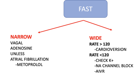
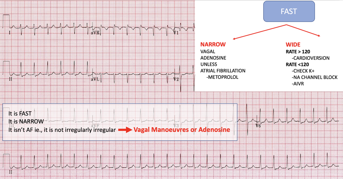
Below is the fast portion of the algorithm and a couple of examples from the course to try.
Picture
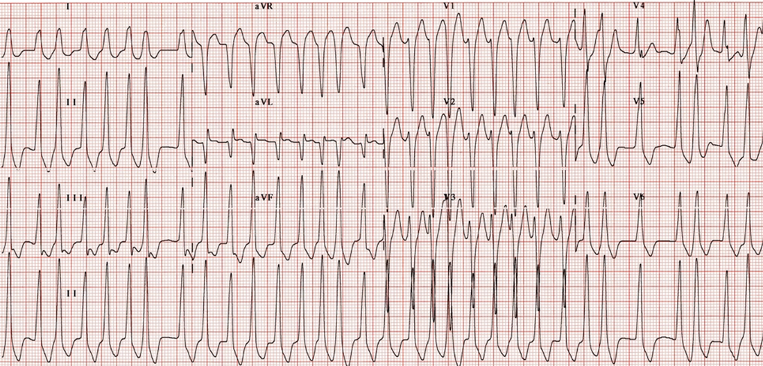
What does the ecg below show?
Picture
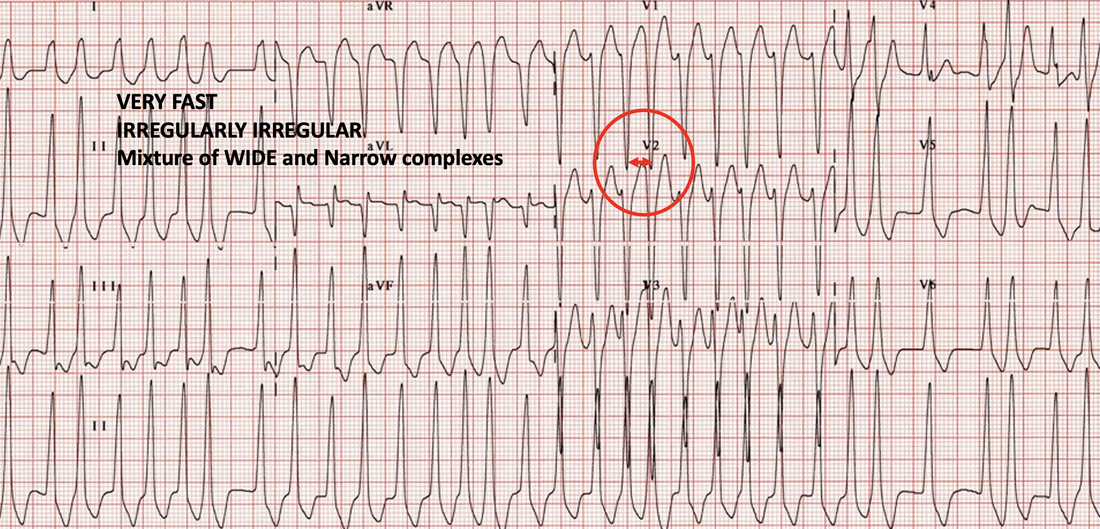
This is WPW with Atrial Fibrillation. The rate in some areas is 300 bpm. If you have any rate faster than 250bpm in an adult, there must be an accessory pathway. It is irregularly irregular. Some complexes are wide and others are narrow, as some pass through the AV node and others via the accessory pathway. See below:
Picture
How do we manage this condition?
If we used Procainamide, that would be fine, as it preferentially blocks the accessory pathway. What we cannot use, is an AV nodal blocker, such as a beta or calcium channel blocker, or Amiodarone. If we did this all the impulses would be conducted down the accessory pathway and the heart would go into ventricular fibrillation.

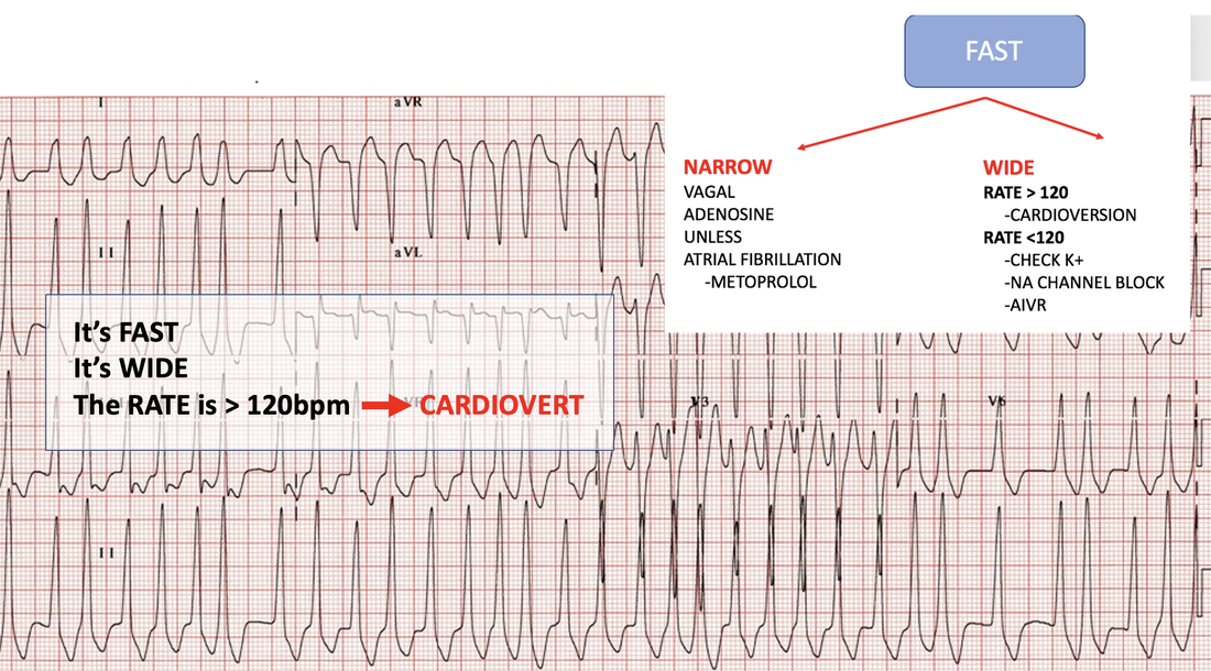
What if we applied the simple algorithm approach?
​
We don't need to know what the diagnosis is.... that's the benefit of having an approach like this. Just follow the flow....
Picture
Let's try one more, then you can go to the course and do a bundle of examples.
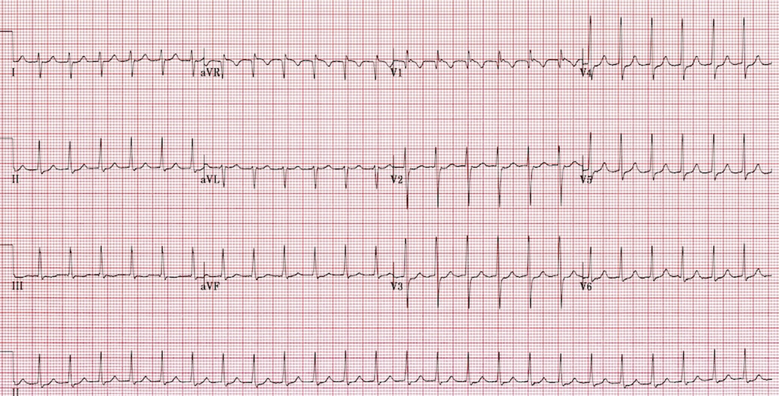
What is the diagnosis of the ECG below?
Picture
It is a fast, narrow complex tachycardia. It is AVN re-entry tachycardia. What if you weren't sure of what to do? What would the algorithm say?
Picture
Remember to develop your own approach or just use mine. It's very satisfying when you have a simple approach. You can then add all the other learnings to this.

​Join me for an a fun approach to really understanding  arrhythmias, when I cover 'Rule the Arrhythmia'  at the Cardiac Bootcamp on the Gold Coast  on January 18 2022, 
0 Comments

    Author

    Dr Peter Kas
    ​Emergency Physician

    Picture
    Picture
    Sign up for
    ECG of the week
     Its free

    Archives

    February 2023
    January 2023
    December 2022
    November 2022
    October 2022
    September 2022
    August 2022
    July 2022
    June 2022
    March 2022
    December 2021
    November 2021
    October 2021
    September 2021
    August 2021
    July 2021
    June 2021
    May 2021
    April 2021
    March 2021
    February 2021
    January 2021
    December 2020
    November 2020
    September 2020
    August 2020
    July 2020

    Categories

    All
    Atrial Fibrillation
    Atrial Flutter
    Atrial Tachycardia
    Axis
    Bradycardia
    Cardiac Arrest
    Chest Pain
    ECG Mimics
    Ischaemia
    Lead Reversal
    Myopericarditis
    Pacing
    Paediatric Arrhythmias
    Paediatric ECG
    Paediatric Syncope
    Pericarditis
    QT Interval
    STEMI
    STEMI Mimics
    SVT
    Ventricular Tachycardia
    Wellens
    Wide Complex Tachycardia

Our Other Websites to Visit

www.emcore.com.au
www.resus.com.au

Contact Us
© COPYRIGHT 2015. ALL RIGHTS RESERVED.
  • HOME
  • Course Login
    • Log In
    • Log Out
  • SYLLABUS
  • STORE
    • Online Self Study Course
    • Cardiac Video Course
  • ECG OF THE WEEK