CARDIAC BOOTCAMP
  • HOME
  • Course Login
    • Log In
    • Log Out
  • SYLLABUS
  • STORE
    • Online Self Study Course
    • Cardiac Video Course
  • ECG OF THE WEEK

ECG OF THE WEEK

BECOME A COURSE MEMBER
Each Week we put up one ECG case for you....because it's easier to learn from cases.
The ECG of the week is FREE to everyone. To become an expert at ECG's join the Cardiac Bootcamp Course. It's a great way to Learn!
​Or come to one of our LIVE Cardiac Bootcamps

the vt score: 100% specificity for vt

14/12/2021

0 Comments

 
Introduction
When we assume that every wide complex tachycardia is Ventricular Tachycardia(VT) our sensitivity for picking up VT is 100%. In the emergency department setting, this is a must! We use electricity on all of these patients because it is safe and effective. We can try adenosine, in case it’s a supra ventricular tachycardia(SVT) but only if it’s a regular rhythm.
Our specificity in this scenario lags, however in our favour, is that fact that 80% of WCT is VT. If we wanted to determine if an ECG is showing VT, we have a range of rules at our disposal,  including Brugada and Vereckei, that give a respectable specificity, but it’s still in the mid 80%.
A new validated rule
The VT Score, uses 7 criteria, some of which we already use, to diagnose VT.  A specificity of 100% is reached when 4 or more criteria are satisfied(1).
Picture
The Seven Criteria are as follows:
NOTE: Before we start I would add that the rate should be greater than 120 beats per minute for us to start considering VT.
1. Dominant Initial R wave in V1. This can be notched(the original rabbit’s ear) provided the notch is on the descending limb.
Picture
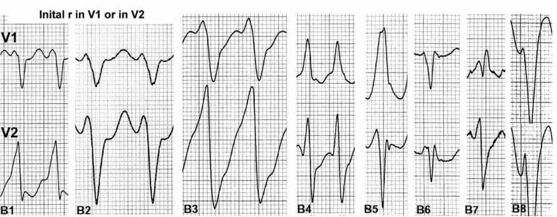
2. Initial r > 40 ms in V1 or V2: assess in predominantly negative QRS complexes. Look for a ‘fat’ initial r in rS in V1. It can also include variations such as: RS with high ‘r’ amplitude, rS with notched r
Picture
3 Notched S in V1. The notch can be at the beginning, in the middle or near the nadir of the S wave.(This has been known as Josephson’s Sign)
Picture
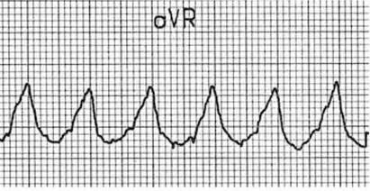
4. Initial R wave in aVR. The QRS in aVR must start with a large R wave. it can be notched. (This comes from the Vereckei(2) criteria).
Picture
5. Lead II R wave Peak time(RWPT) > or equal to 50ms. This is the time from the beginning of QRS to first change in direction.
Picture
6. Lack of RS in V1-V6.(Brugada Criterion(3)). This is similar to saying that there is concordance.
Picture
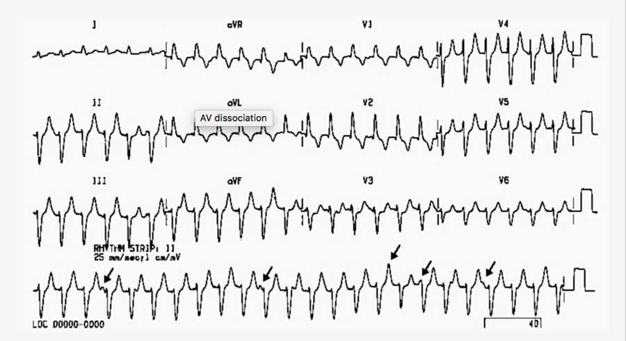
7. Atrioventricular Dissociation. This is indicated where a fast ventricular activity iOS not related to atrial depolarisation.
Picture
Source: LITFL
LETS MAKE IT EASIER TO REMEMBER:
  1. Rate must be > 120bpm to be VT
  2. Concordance in V1-V6
  3. AV Dissociation
  4. Josephson’s Sign in V1 -V2
  5. Look at V1 for a FAT INITIAL R wave (r>40ms) this could be the rabbits ear
  6. Look at aVR for a FAT Wave to begin with
  7. Look at lead II RWPT > 50ms- again a big R wave.
References
  1. Jastrzebski M et al. The ventricular tachycardia score: a novel approach to electrocardiographic diagnosis of ventricular tachycardia. Europace. 2016 Apr;18(4):578-84
  2. Vereckei A et al. Application of a new algorithm in the differential diagnosis of wide QRS complex tachycardia. Eur Heart J 2007.28:589-600
  3. Brugada P. et al. A new approach to the differential diagnosis of a regular tachycardia with a wide QRS complex. Circulation 1991:83:1649-1659
0 Comments


Leave a Reply.

    Author

    Dr Peter Kas
    ​Emergency Physician

    Back to resus.com.au
    Picture
    Picture
    Sign up for
    ECG of the week
     Its free

    Archives

    May 2023
    March 2023
    February 2023
    January 2023
    December 2022
    November 2022
    October 2022
    September 2022
    August 2022
    July 2022
    June 2022
    March 2022
    December 2021
    November 2021
    October 2021
    September 2021
    August 2021
    July 2021
    June 2021
    May 2021
    April 2021
    March 2021
    February 2021
    January 2021
    December 2020
    November 2020
    September 2020
    August 2020
    July 2020

    Categories

    All
    Ablation
    Atrial Fibrillation
    Atrial Flutter
    Atrial Tachycardia
    Axis
    Bradycardia
    Cardiac Arrest
    Chest Pain
    ECG Mimics
    Ischaemia
    Lead Reversal
    Migraine
    Myopericarditis
    Pacing
    Paediatric Arrhythmias
    Paediatric ECG
    Paediatric Syncope
    Pericarditis
    QT Interval
    STEMI
    STEMI Mimics
    SVT
    T Waves
    Ventricular Tachycardia
    Wellens
    Wide Complex Tachycardia

Our Other Websites to Visit

www.emcore.com.au
www.resus.com.au

Contact Us
© COPYRIGHT 2015. ALL RIGHTS RESERVED.
  • HOME
  • Course Login
    • Log In
    • Log Out
  • SYLLABUS
  • STORE
    • Online Self Study Course
    • Cardiac Video Course
  • ECG OF THE WEEK