CARDIAC BOOTCAMP
  • HOME
  • Course Login
    • Log In
    • Log Out
  • SYLLABUS
  • STORE
    • Online Self Study Course
    • Cardiac Video Course
  • ECG OF THE WEEK

ECG OF THE WEEK

BECOME A COURSE MEMBER
Each Week we put up one ECG case for you....because it's easier to learn from cases.
The ECG of the week is FREE to everyone. To become an expert at ECG's join the Cardiac Bootcamp Course. It's a great way to Learn!
​Or come to one of our LIVE Cardiac Bootcamps

A 38 yo male presents with chest pain

2/7/2022

1 Comment

 
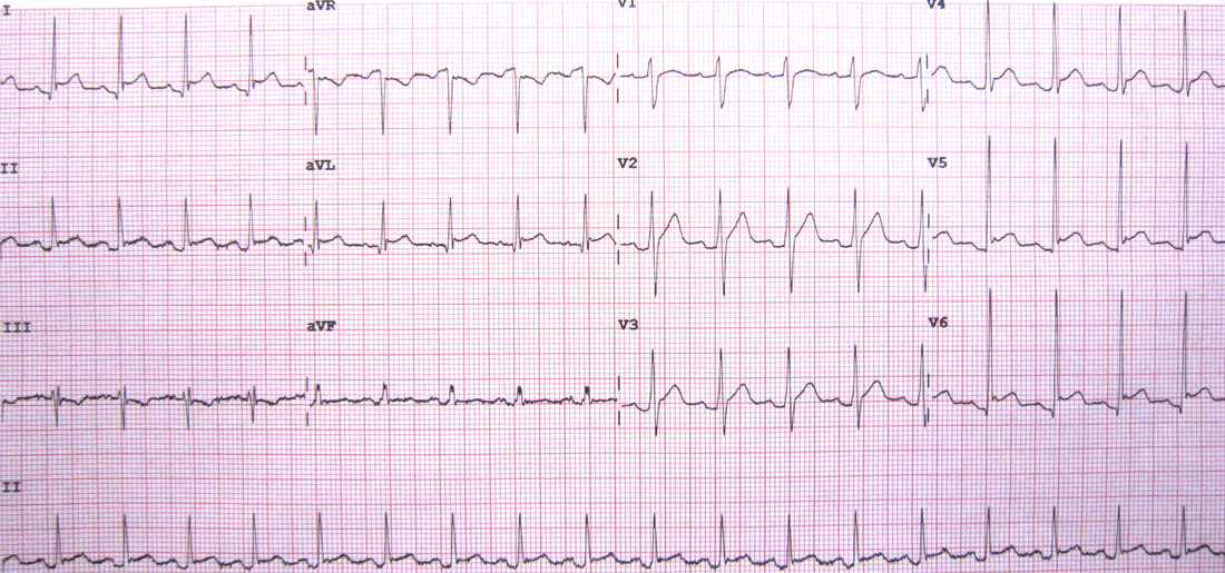
A 38 yo male presents with sharp stabbing central chest pain. It changes with breathing. He feels worst when he is lying down. He is haemodynamically stable. An ECG is done. What does it show?
Picture
WHAT DOES THE ECG SHOW?
Using the ECG in 20 Seconds Approach:
  • There is a sinus tachycardia at about 108bpm
    • The P wave is upright in II and inverted in aVR, so it is sinus
  • The QRS morphology is normal
  • The ST-T segments are not normal:
    • There is ST elevation in multiple leads ie., I, II aVL, V4, V5, V6
    • There appears to be a J point in aVL, V4, V5 and V6, so Benign early repolarisation is an alternative diagnosis.
    • There is ST depression in aVR
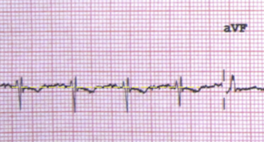
    • Is that ST depression in III?​
      • Initially it does look like ST depression, however when we draw in the isoelectric line, it is not really as significant (see below).
Picture
REMEMBER: When looking for ST elevation or depression, use the T-P line as the isoelectric line.
  • The QT and PR intervals are normal
    • It is obvious that there is PR depression
  • There are no pacing spikes
DIAGNOSIS
The diagnosis is most likely pericarditis.
The history is very indicative of that. The characteristics of the pain ie., that is it worst on lying down indicate pericarditis. Although there appears to be a pleuritic component, there is no real other history of a PE and the ECG does not show a right axis or S1Q3T3 etc.

In the ECG itself:
  • There is ST elevation, concave in nature
  • There is ST depression in aVR, which is allowed ie., there can be ST depression in aVR and V1
  • There are no ischaemic looking waveforms and no real reciprocal changes.
  • There is PR depression.
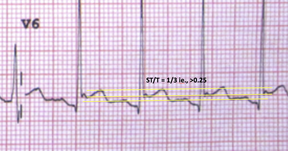
  • We can also measure the ST/T ratio. A ratio of >0.25, when put together with all the other parameters indicates pericarditis.
Picture
WHAT TO DO NEXT
  • Do a bedside echo looking for an effusion. It will also assist to look for wall abnormalities.
  • Get a troponin. A slight rise in troponin may be normal. We need to make sure that we don't miss, perimyocarditis or myocarditis, where we would expect a significant rise in troponin.
Who needs admission and who can be discharged?
What is Spodick's Sign and how good is it?
What are the stages of Pericarditis?
What is the treatment?
​Is there a role for glucocorticoids?
Members login and go to the pericarditis section for answers to all the above and more
1 Comment
Marin
5/7/2022 05:18:07 pm

Hi Peter,

Thank you very much for this interesting and always challenging ECG.
Peter, you did not mention an obvious PR elevation in aVR as one of the ECG features of Pericarditis.
Was this just simple overlooking of something that we always look for or PR elevation in aVR is not relevant.
Cheers
Marin

Reply


Leave a Reply.

    Author

    Dr Peter Kas
    ​Emergency Physician

    Picture
    Picture
    Sign up for
    ECG of the week
     Its free

    Archives

    February 2023
    January 2023
    December 2022
    November 2022
    October 2022
    September 2022
    August 2022
    July 2022
    June 2022
    March 2022
    December 2021
    November 2021
    October 2021
    September 2021
    August 2021
    July 2021
    June 2021
    May 2021
    April 2021
    March 2021
    February 2021
    January 2021
    December 2020
    November 2020
    September 2020
    August 2020
    July 2020

    Categories

    All
    Atrial Fibrillation
    Atrial Flutter
    Atrial Tachycardia
    Axis
    Bradycardia
    Cardiac Arrest
    Chest Pain
    ECG Mimics
    Ischaemia
    Lead Reversal
    Myopericarditis
    Pacing
    Paediatric Arrhythmias
    Paediatric ECG
    Paediatric Syncope
    Pericarditis
    QT Interval
    STEMI
    STEMI Mimics
    SVT
    Ventricular Tachycardia
    Wellens
    Wide Complex Tachycardia

Our Other Websites to Visit

www.emcore.com.au
www.resus.com.au

Contact Us
© COPYRIGHT 2015. ALL RIGHTS RESERVED.
  • HOME
  • Course Login
    • Log In
    • Log Out
  • SYLLABUS
  • STORE
    • Online Self Study Course
    • Cardiac Video Course
  • ECG OF THE WEEK