CARDIAC BOOTCAMP
  • HOME
  • Course Login
    • Log In
    • Log Out
  • SYLLABUS
  • STORE
    • Online Self Study Course
    • Cardiac Video Course
  • ECG OF THE WEEK

ECG OF THE WEEK

BECOME A COURSE MEMBER
Each Week we put up one ECG case for you....because it's easier to learn from cases.
The ECG of the week is FREE to everyone. To become an expert at ECG's join the Cardiac Bootcamp Course. It's a great way to Learn!
​Or come to one of our LIVE Cardiac Bootcamps

Beware of Imposter Arrhythmias

29/7/2020

0 Comments

 
In the Wide Complex Rhythms part of the Course, I talk about the mimics of VT. However that's not all that can be mimicked.
If we don't recognised the mimic, we can actually do harm to the patient by giving treatment that isn't needed.
​
CASE 1
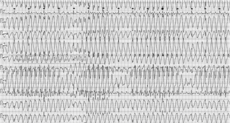
​A holter monitor is done on a young patient who has complained of palpitations. His tracing is shown.
A diagnosis of VT is made. It's easy to be tricked. See the second ecg below.
Picture
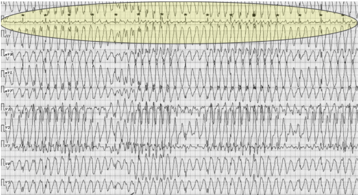
Look at lead I below. It is not showing a wide complex. You can't have one lead refusing to show VT and all the others doing so. This is not a wide complex. This tracing was taken during a time when the patient was playing a video game on their phone. The tapping came produced this complex.
Picture
A similar interference pattern can occur secondary to tremor, or rigors, or someone just tapping on the lead.
Below is another case.

​CASE 2
A 35 yo male with known Cardiomyopathy presents to the ED with chest pain.
The diagnosis of atrial flutter is made by the registrar, after seeing the rhythm on the monitor.
He is planning for definitive treatment; in this case cardioversion. That's another discussion. (I wouldn't necessarily cardiovert atrial flutter on presentation.)
An ECG is done.
Is this atrial flutter?
What else could it be if it isn't flutter?

​See the ECG below:
Picture
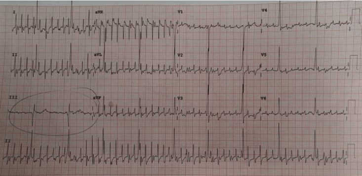
This is nothing more than artefact. If you look closely at the ECG you can see that the patient’s underlying rhythm. The tall QRS complexes (well seen in V4-6), are the patient's underlying rhythm. There is left ventricular hypertrophy due to the patient’s cardiomyopathy. The waveforms in-between these, are simply artefact, just noise. In this case they are created due to the patients underlying tremor.
The 12 lead ECG was seen and the diagnosis of flutter was still entertained by the registrar.
If we look at this ECG, we can see where I’ve circled the complexes in lead III.
You can’t have atrial flutter in every lead, but lead III refuses to go into flutter. It’s artefact created by patient tremor and movement. It was easily reproduced by my tapping on the leads.

Now even though this wasn’t flutter we still needed to sort out the chest pain with serial Troponins.
0 Comments


Leave a Reply.

    Author

    Dr Peter Kas
    ​Emergency Physician

    Back to resus.com.au
    Picture
    Picture
    Sign up for
    ECG of the week
     Its free

    Archives

    May 2023
    March 2023
    February 2023
    January 2023
    December 2022
    November 2022
    October 2022
    September 2022
    August 2022
    July 2022
    June 2022
    March 2022
    December 2021
    November 2021
    October 2021
    September 2021
    August 2021
    July 2021
    June 2021
    May 2021
    April 2021
    March 2021
    February 2021
    January 2021
    December 2020
    November 2020
    September 2020
    August 2020
    July 2020

    Categories

    All
    Ablation
    Atrial Fibrillation
    Atrial Flutter
    Atrial Tachycardia
    Axis
    Bradycardia
    Cardiac Arrest
    Chest Pain
    ECG Mimics
    Ischaemia
    Lead Reversal
    Migraine
    Myopericarditis
    Pacing
    Paediatric Arrhythmias
    Paediatric ECG
    Paediatric Syncope
    Pericarditis
    QT Interval
    STEMI
    STEMI Mimics
    SVT
    T Waves
    Ventricular Tachycardia
    Wellens
    Wide Complex Tachycardia

Our Other Websites to Visit

www.emcore.com.au
www.resus.com.au

Contact Us
© COPYRIGHT 2015. ALL RIGHTS RESERVED.
  • HOME
  • Course Login
    • Log In
    • Log Out
  • SYLLABUS
  • STORE
    • Online Self Study Course
    • Cardiac Video Course
  • ECG OF THE WEEK