CARDIAC BOOTCAMP
  • HOME
  • Course Login
    • Log In
    • Log Out
  • SYLLABUS
  • STORE
    • Online Self Study Course
    • Cardiac Video Course
  • ECG OF THE WEEK

ECG OF THE WEEK

BECOME A COURSE MEMBER
Each Week we put up one ECG case for you....because it's easier to learn from cases.
The ECG of the week is FREE to everyone. To become an expert at ECG's join the Cardiac Bootcamp Course. It's a great way to Learn!
​Or come to one of our LIVE Cardiac Bootcamps

Atrial fibrillation: HOW would you treat?

22/6/2022

0 Comments


Case 1

​A 68 yo male presents with palpitations and vague chest tightness. He has noticed his heart racing for the last 2 hours. His ECG shows atrial fibrillation, with a rapid ventricular response.  He has a past medical history of hypertension and smoking.
He is afebrile, with a heart rate of 176-185 beats per minute, BP of 138/65, Sats of 96% on room air.
How would you manage this patient's atrial fibrillation?

Case 2

​​A 62 yo male presents with palpitations and vague chest discomfort. He has noticed his heart racing for the last 2 hours. His ECG shows atrial fibrillation, with a rapid ventricular response. He has a past medical history of hypertension and smoking. He is febrile eat 38.3 C, a heart rate of 176-185 beats per minute, BP of 138/65, Sats of 94% on room air. On clinical examination, has midzone crepitations in his right lung.
​How would you manage this patient's atrial fibrillation and would it be different to the patient in case 1?

Deciding on the treatment of atrial fibrillation. 

It’s all about primary vs secondary atrial fibrillation/flutter.
A recent review of the approach to the Emergency Department(ED) patient with atrial fibrillation/flutterwas published by the Canadian Association of Emergency Physicians(1).
Here is a synopsis of the approach.

Is it PRIMARY or SECONDARY?

Is there an underlying disease causing the atrial fibrillation/flutter(AF/Flut), or it is a primary event?
Primary AF/Flut is usually of sudden onset and is not due to an underlying medical condition causing. Secondary AF/Flut is usually due to a medical cause such as sepsis or pulmonary embolism or bleeding or acute coronary syndrome. Secondary causes are usually not sudden in onset and tend to have slower ventricular rates(<150), but not always.
Why is this important?
We can aggressively treat Primary AF/Flut, however we need to treat the cause of secondary AF/Flut, not the arrhythmia itself, as aggressive rate or rhythm control in secondary causes can be harmful.

PRIMARY AF/FLUT
We need to ensure that the patient is haemodynamically stable.
An unstable patient may show the following:
  • Systolic Blood Pressure <90 mmHg or signs of shock(altered mental state)
  • Signs of cardiac ischaemia: chest pain, ST depression on ECG
  • Pulmonary Oedema
The unstable patient needs electrical cardio version if the arrhythmia has been present for < 48hours, or in those where it has been there for >48hours, rate control.

In the stable patient, when is it safe to cardiovert Primary AF/Flut?
Rhythm control is always preferred to rate control.
In this article it was considered safe to cardiovert if:
The patient was anticoagulated for > 3 weeks
OR
The patient is not anticoagulated for > 3 weeks but 
Has no history of TIA or stroke 
Has no valvular heart disease  and
  • Onset < 12 hours ago OR
  • Onset 12-48 hours ago and < 2 CHADS-65 Criteria 
    • (age >65, Diabetes, HT, heart failure Stroke or TIA- see below) OR
  • No thrombus on TOE

RATE Control
Rate Control is used when cardioversion (rhythm control) is unsafe. Our target is a heart rate < 100.
If the patient is already on a beta blocker or a calcium channel blocker, more of the same medication may be given.
  • Beta Blockers: 
    • Metoprolol
      • 2.5-5mg IV over 2 min. 
      • Repeat q15-20min for 3 doses. 
      • Commence 25-50 mg PO  within 30 minutes of IV control. 
      • Discharge on 25-50mg bd. 
  • Calcium Channel Blockers
    • Diltiazem
      • Avoid these in acute heart failure or known LV dysfunction
      • Diltiazem 0.25 mg/kg IV over 10 min repeat q15-20 min 3 doses
  • Digoxin
    • Second line, slow onset- BUT FIRST LINE if hypotension and acute heart failure
      • 0.25-0.5mg loading dose then0.25mg IV Q 4-6 hours to max 1.5mg over 24 hours
      • BEWARE RENAL FAILURE
In a recent study in the American Journal of Emergency Medicine(2), “Intravenous diltiazem has higher efficacy, shorter average onset time, lower ventricular rate, less impact on blood pressure, and with no increase in adverse events compared to intravenous metoprolol.” Unfortunately IV Diltiazem is not readily available in Australia.

RHYTHM Control
Electrical Cardioversion
  • Pads – antero-lateral or antero-posterior
    • In a recent multicenter study in Circulation(3) it was found that anterolateral positioning was more effective for biphasic cardioversion of atrial fibrillation
  • Start with 150-200J avoid low energy. AF is one of the most resistant arrhythmias, so start high.
  • Note that pre-treatment with an anti arrhythmic is not recommended.
Pharmacological Cardioversion
  • Procainamide 
    • IV 15mg/kg in 500ml NSaline over 60min. Maximum 1500mg
    • Beware:•Hypotension- do not use if SBP<100mmHg 
    • Stop the infusion if 
      • BP drops
      • Long QTc- Don’t use if QTc is >500ms
      • Stop if QTc lengthens and Check QTc after cardioversion•
  • Amiodarone– NOT recommended: It has slow onset and low efficacy

STROKE PREVENTION
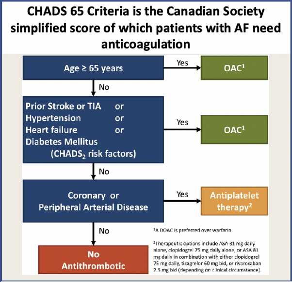
Who needs anticoagulation?
​CHADS-65 POSITIVE (age >65, DM, HT, CCF, Stroke/TIA)
Picture
​
  • DOAC preferred
  • WARFARIN MUST BE USED in
    • Mechanical Valve
    • Moderate to severe Mitral Stenosis
    • Severe renal Impairment CrCl <30ml/min
  • What if the patient is on ASPIRIN?
    • If stable CAD- stop aspirin
    • If CAD with other antiplatelets OR PCI<12 months D/W Cardiology
CHADS-65 NEGATIVE
  • Consider OAC for 4 weeks, but needs shared decision making with the patient.
  • If stable CAD patient can continue aspirin
If planning Trans-oesophageal Echo guided cardioversion: Anticoagulate for 4 weeks
Remember that even if the patient reverts spontaneously, anticoagulation should still be given if there are risk factors.

Anticoagulation Checklist
  • Dabigatran 150mg BD: 110mg bd if >80 or >75 with bleed risk
  • Rivaroxaban 20mg daily: 15mg daily if CrCl 30-49ml/min
  • Apixaban 5mg BD: 2.5mg bd if 2 of: Creat >133umol/L, Age > 80yo, Weight <60kg. Otherwise 5mg bd
  • Edoxaban 60mg daily: 30mg if CrCl 30-50ml/min or wt<60kg
  • Warfarin start 5mg daily: 1-2mg if frail, low weight or Asian descent, INR after 3-4 doses of Warfarin
Who to Admit
  • Symptomatic despite treatment
  • Have ACS and chest pain and ECG changes
  • Acute heart failure
  • NOTE: in uncomplicated Afib/Flut, admission is rarely needed
  • Expect a Troponin leak

References
  1. Still I.G. et al. 2021 CAEP Acute Atrial Fibrillation/Flutter Best Practices Checklist. Canadian Journal of Emergency Medicine (2021) 23:604-610.
  2. Lan Q et al. Intravenous Diltiazem versus metoprolol for atrial fibrillation and rapid ventricular rate: A meta-analysis. The American Journal of Emergency Medicine. Vol51: (Jan 2022); 248-256
  3. Schmidt A s et al. Anterior-Lateral versus Anterior-Posterior Electrode Position for Cardioverting Atrial Fibrillation. Circulation (2021); 144:1995-2003
Members go to the atrial fibrillation section for a discussion of the detailed approach
0 Comments


Leave a Reply.

    Author

    Dr Peter Kas
    ​Emergency Physician

    Picture
    Picture
    Sign up for
    ECG of the week
     Its free

    Archives

    February 2023
    January 2023
    December 2022
    November 2022
    October 2022
    September 2022
    August 2022
    July 2022
    June 2022
    March 2022
    December 2021
    November 2021
    October 2021
    September 2021
    August 2021
    July 2021
    June 2021
    May 2021
    April 2021
    March 2021
    February 2021
    January 2021
    December 2020
    November 2020
    September 2020
    August 2020
    July 2020

    Categories

    All
    Atrial Fibrillation
    Atrial Flutter
    Atrial Tachycardia
    Axis
    Bradycardia
    Cardiac Arrest
    Chest Pain
    ECG Mimics
    Ischaemia
    Lead Reversal
    Myopericarditis
    Pacing
    Paediatric Arrhythmias
    Paediatric ECG
    Paediatric Syncope
    Pericarditis
    QT Interval
    STEMI
    STEMI Mimics
    SVT
    Ventricular Tachycardia
    Wellens
    Wide Complex Tachycardia

Our Other Websites to Visit

www.emcore.com.au
www.resus.com.au

Contact Us
© COPYRIGHT 2015. ALL RIGHTS RESERVED.
  • HOME
  • Course Login
    • Log In
    • Log Out
  • SYLLABUS
  • STORE
    • Online Self Study Course
    • Cardiac Video Course
  • ECG OF THE WEEK